. 2019 Jan 24;9(1):471.
doi: 10.1038/s41598-018-36712-6.

Initiation of Pulmonary Fibrosis after Silica Inhalation in Rats is linked with Dysfunctional Shelterin Complex and DNA Damage Response

Affiliations

Initiation of Pulmonary Fibrosis after Silica Inhalation in Rats is linked with Dysfunctional Shelterin Complex and DNA Damage Response

Mohammad Shoeb et al. Sci Rep. .

Abstract

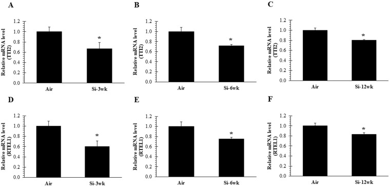
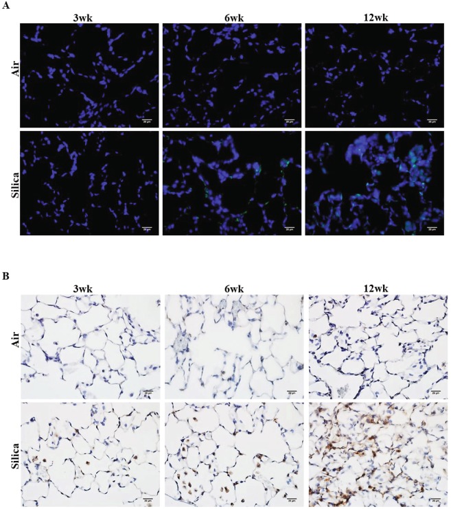
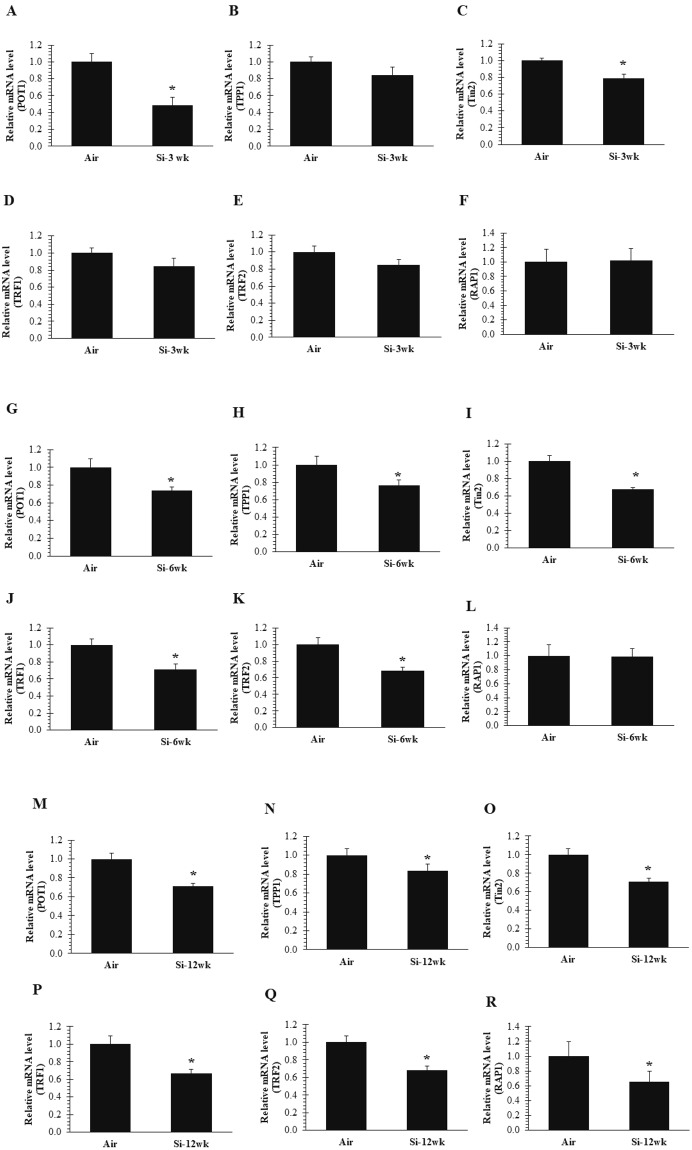
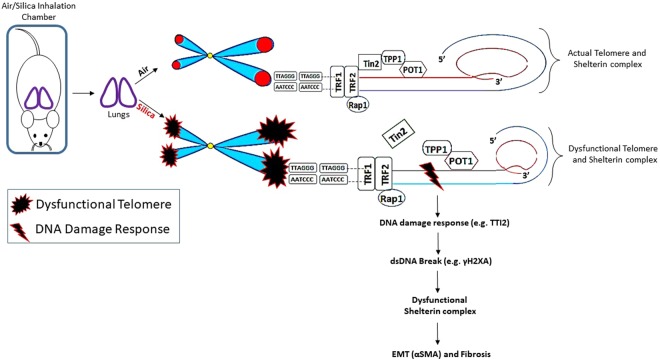
Occupational exposure to silica has been observed to cause pulmonary fibrosis and lung cancer through complex mechanisms. Telomeres, the nucleoprotein structures with repetitive (TTAGGG) sequences at the end of chromosomes, are a molecular "clock of life", and alterations are associated with chronic disease. The shelterin complex (POT1, TRF1, TRF2, Tin2, Rap1, and POT1 and TPP1) plays an important role in maintaining telomere length and integrity, and any alteration in telomeres may activate DNA damage response (DDR) machinery resulting in telomere attrition. The goal of this study was to assess the effect of silica exposure on the regulation of the shelterin complex in an animal model. Male Fisher 344 rats were exposed by inhalation to Min-U-Sil 5 silica for 3, 6, or 12 wk at a concentration of 15 mg/m3 for 6 hr/d for 5 consecutive d/wk. Expression of shelterin complex genes was assessed in the lungs at 16 hr after the end of each exposure. Also, the relationship between increased DNA damage protein (γH2AX) and expression of silica-induced fibrotic marker, αSMA, was evaluated. Our findings reveal new information about the dysregulation of shelterin complex after silica inhalation in rats, and how this pathway may lead to the initiation of silica-induced pulmonary fibrosis.


Conflict of interest statement

The authors declare no competing interests.

Figures

Figure 1
Figure 1
Inactivation of TTI2 and RTEL1 after 3, 6, and 12 wk of silica inhalation. (A–C) TTI2 and (D–F) RTEL1 expression in lung cDNA at 16 hr after a 3, 6, and 12 wk silica inhalation exposure (15 mg/m3, 6 h/d, 5 d/wk). Rats exposed to filtered air served as the controls; n = 8; values are means ± standard error; *significantly different from corresponding air control, p < 0.05.
Figure 2
Figure 2
Upregulation of expression of γH2AX and αSMA after 3, 6, and 12 wk of silica inhalation. (A) Representative image of γH2AX positive cells (green), and (B) αSMA expression (brown) in lung tissue at 16 hr after a 3, 6, and 12 wk silica inhalation exposure (15 mg/m3, 6 h/d, 5 d/wk). The scale bar denotes 20 µm. Rats exposed to filtered air served as the controls; n = 5.
Figure 3
Figure 3
Disruption of only Tin2 of the shelterin core complex after 3 wk of silica inhalation. Inactivation of (A) POT1 and (C) Tin2 expression in lung cDNA at 16 hr after a 3-wk silica inhalation exposure (15 mg/m3, 6 h/d, 5 d/wk, 3 wk). (B) TPP1 (D) TRF1 (E) TRF2 and (F) RAP1 remained unchanged. All shelterin components expression (G) POT1 (H) TPP1 (I) Tin2 (J) TRF1 (K) TRF2 except (L) RAP1 were significantly downregulated in lung cDNA at 16 hr after a 6-wk silica inhalation exposure (15 mg/m3, 6 h/d, 5 d/wk, 6 wk). All shelterin components expression (M) POT1 (N) TPP1 (O) Tin2 (P) TRF1 (Q) TRF2 and (R) RAP1 were significantly downregulated in lung cDNA at 16 hr after a 12-wk silica inhalation exposure (15 mg/m3, 6 h/d, 5 d/wk, 12 wk). Rats exposed to filtered air served as the controls; n = 8; values are means ± standard error; *significantly different from corresponding air control, p < 0.05.
Figure 4
Figure 4
Schematic diagram of the initiation of pulmonary fibrosis after silica inhalation is associated with shelterin dysregulation and dsDNA break in rat lung that may lead to a persistent and severe DDR (TTI2) at telomeres characterized by increased γH2AX and αSMA. Silica inhalation caused an increased staining in the number of γH2AX positive cells and an elevated αSMA expression in lung tissue compared to air control. Initiation of shelterin dysregulation was started after 3 wk of silica exposure as shown by the disruption of Tin2-TPP1 and remained persistent at 6 and 12 wk of silica exposure.

Similar articles

Cited by

References

    1. Centers for Disease Control and Prevention. NIOSH Workplace Safety and Health Topic- Silica. http://www.cdc.gov/niosh/topics/silica/default.html (2017).
    1. King TE, Jr., Pardo A, Selman M. Idiopathic pulmonary fibrosis. Lancet. 2011;378:1949–1961. doi: 10.1016/S0140-6736(11)60052-4. - DOI - PubMed
    1. Selman M, King TE, Pardo A. American Thoracic Society; European Respiratory Society; American College of Chest Physicians. Idiopathic pulmonary fibrosis: prevailing and evolving hypotheses about its pathogenesis and implications for therapy. Ann. Intern. Med. 2001;134:136–151. doi: 10.7326/0003-4819-134-2-200101160-00015. - DOI - PubMed
    1. Hinz B, et al. The myofibroblast: one function, multiple origins. Am. J. Pathol. 2007;170:1807–1816. doi: 10.2353/ajpath.2007.070112. - DOI - PMC - PubMed
    1. Araya, J. & Nishimura, S. L. Fibrogenic reactions in lung disease. Annu Rev Pathol 5, 77–98, https://doi.org/10.1146/annurev.pathol.4.110807.092217 (2010). - PubMed

Publication types