. 2019 Jan 24;9(1):721.
doi: 10.1038/s41598-018-36809-y.

Open-Source Multiparametric Optocardiography

Affiliations

Open-Source Multiparametric Optocardiography

Brianna Cathey et al. Sci Rep. .

Abstract

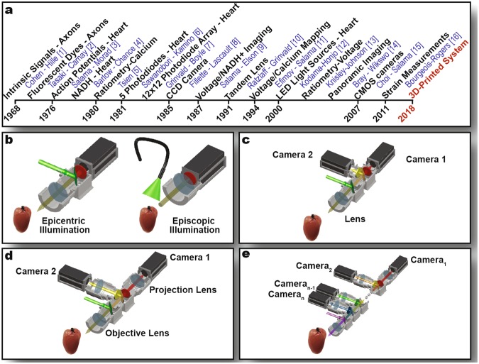
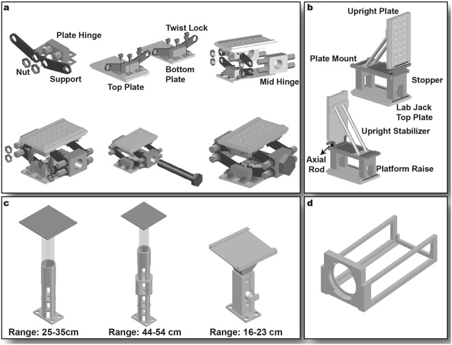
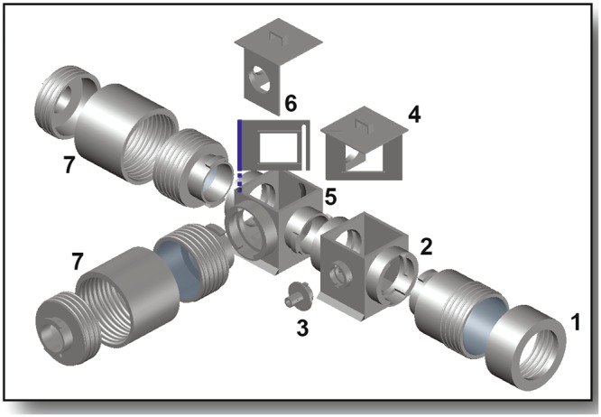
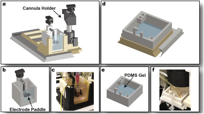
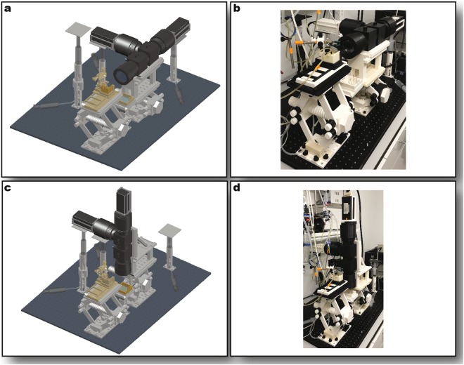
Since the 1970s fluorescence imaging has become a leading tool in the discovery of mechanisms of cardiac function and arrhythmias. Gradual improvements in fluorescent probes and multi-camera technology have increased the power of optical mapping and made a major impact on the field of cardiac electrophysiology. Tandem-lens optical mapping systems facilitated simultaneous recording of multiple parameters characterizing cardiac function. However, high cost and technological complexity restricted its proliferation to the wider biological community. We present here, an open-source solution for multiple-camera tandem-lens optical systems for multiparametric mapping of transmembrane potential, intracellular calcium dynamics and other parameters in intact mouse hearts and in rat heart slices. This 3D-printable hardware and Matlab-based RHYTHM 1.2 analysis software are distributed under an MIT open-source license. Rapid prototyping permits the development of inexpensive, customized systems with broad functionality, allowing wider application of this technology outside biomedical engineering laboratories.


Conflict of interest statement

The authors declare no competing interests.

Figures

Figure 1
Figure 1
History of Optical Mapping. (a) Timeline of advances in optical mapping technology. (be) Optical mapping system setups with colored lines representing light path. Single-camera imaging is the simplest implementation. (b) Dual-camera imaging allows the simultaneous measurement of two parameters. (c) Tandem-lens arrangement (d) permits extension of the system for multi-parametric imaging (e).
Figure 2
Figure 2
Stage Components. (a) Lab jack assembly. (b) Tilting platform for upright imaging mode. Emission and excitation filter cubes are mounted onto the upright plate. (c) Lifts. Two hydraulic camera lifts (left and middle) support the cameras in both system orientations. The upright bath lift (right) permits height adjustment of tissue preparation during upright imaging. (d) Camera cage secures cameras to projection lens sleeves.
Figure 3
Figure 3
Optical Components. Expanded view of components housing filters, dichroic mirrors, and lenses (transparent gray). The objective lens sleeve (1) secures the objective lens to the excitation filter cube (2) that guides excitation light to the tissue preparation. The excitation light adaptor (3) secures the excitation light guide to the excitation filter cube. The stationary optics holder (4), placed in the excitation filter cube in the orientation shown, houses a dichroic mirror and features a circular emission filter holder for single-camera imaging. The emission filter cube (5) houses an adjustable wall (6) holding a second dichroic mirror that split the emitted light from the tissue preparation to two cameras (not shown). The projection lens sleeves (7) houses the projection lenses and secures one camera at the end of each.
Figure 4
Figure 4
Perfusion Components. Sideways imaging components. The sideways bath stage (a) houses the sideways bath (b) and an adjacent cannula holder. Pseudo-ECG electrodes fit into the 3 slots of the electrode paddle that also stabilizes the heart against the optical window. (c) Image of printed sideways bath. Upright imaging components. The upright bath stage (d) houses the upright bath (e). PDMS gel secures insect pins holding tissue in place. (f) Image of upright bath.
Figure 5
Figure 5
Full Optical System Assembly. Sideways imaging mode rendering (a) and photo (b). Upright imaging mode rendering (c) and photo (d). In the renderings, stage components are shown in light gray, optical components in dark gray, and perfusion components in gold.
Figure 6
Figure 6
System Demonstration Data. (a) Activation maps of Langendorff-perfused whole mouse heart stained with voltage sensitive dye RH237 (left) and calcium sensitive dye Rhod2AM (right). Representative action potential and calcium transient recordings of whole mouse heart (b) and rat cardiac slice (c) during control Tyrode or 300 µM pinacidil treatment. (d) Comparison of pseudo-ECG traces using electrodes (+, −, gnd) placed in customized electrode paddle (orange) vs. traditional electrode placement (blue). The schematic (f) depicts electrode placement in each case. The graph (e) shows continuous temperature recording of a representative experiment.

References

    1. Cohen LB, Keynes RD, Hille B. Light Scattering and Birefringence Changes during Nerve Activity. Nature. 1968;218:438. doi: 10.1038/218438a0. - DOI - PubMed
    1. Tasaki I, Watanabe A, Sandlin R, Carnay L. Changes in fluorescence, turbidity, and birefringence associated with nerve excitation. Proc. Natl. Acad. Sci. 1968;61:883–888. doi: 10.1073/pnas.61.3.883. - DOI - PMC - PubMed
    1. Salama G, Morad M. Merocyanine 540 as an optical probe of transmembrane electrical activity in the heart. Science. 1976;191:485–487. doi: 10.1126/science.191.4226.485. - DOI - PubMed
    1. Barlow CH, Chance B. Ischemic areas in perfused rat hearts: measurement by NADH fluorescence photography. Science. 1976;193:909–910. doi: 10.1126/science.181843. - DOI - PubMed
    1. Tsien RY. New calcium indicators and buffers with high selectivity against magnesium and protons: design, synthesis, and properties of prototype structures. Biochemistry. 1980;19:2396–2404. doi: 10.1021/bi00552a018. - DOI - PubMed

Publication types