. 2019 Jan 24;9(1):491.
doi: 10.1038/s41598-018-36858-3.

Curcumin inhibits the TGF-β1-dependent differentiation of lung fibroblasts via PPARγ-driven upregulation of cathepsins B and L

Affiliations

Curcumin inhibits the TGF-β1-dependent differentiation of lung fibroblasts via PPARγ-driven upregulation of cathepsins B and L

Ahlame Saidi et al. Sci Rep. .

Abstract

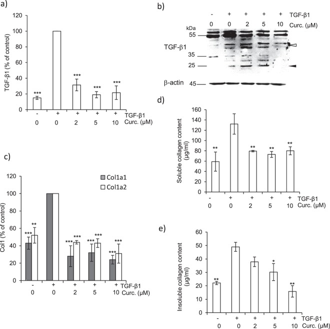
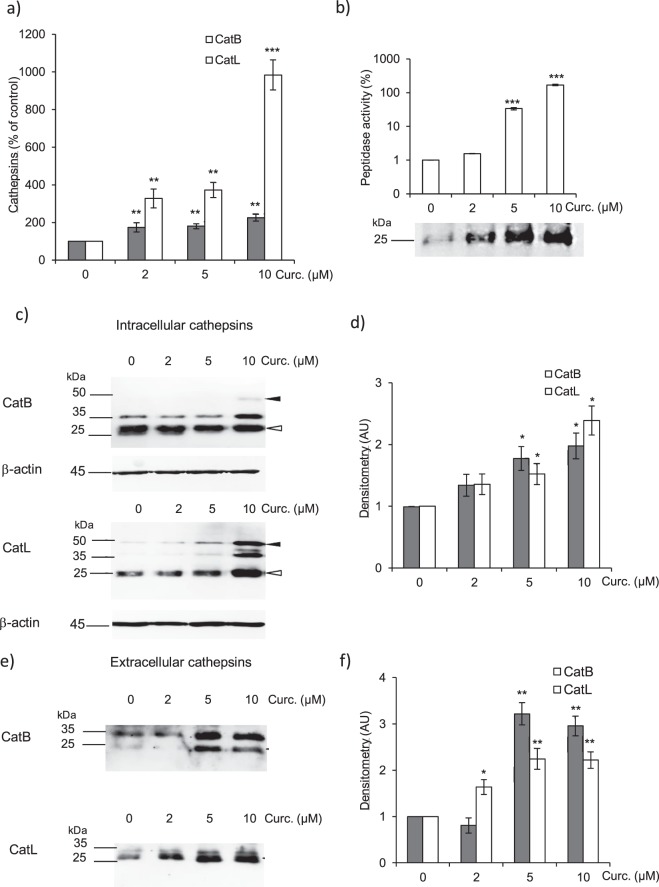
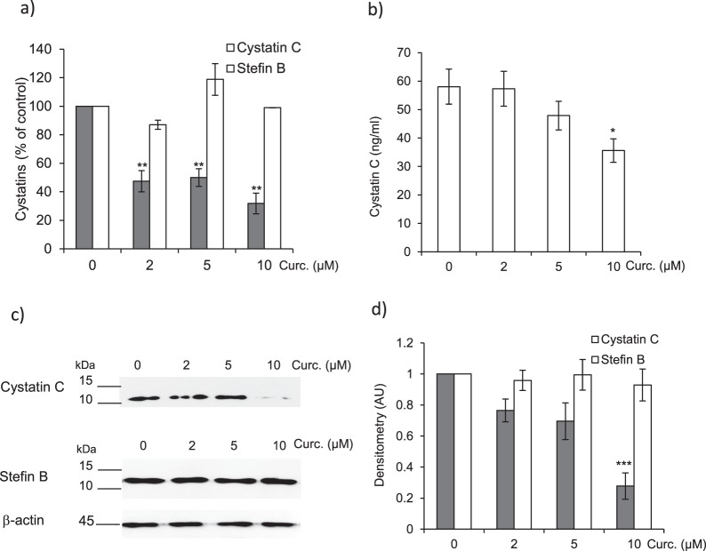
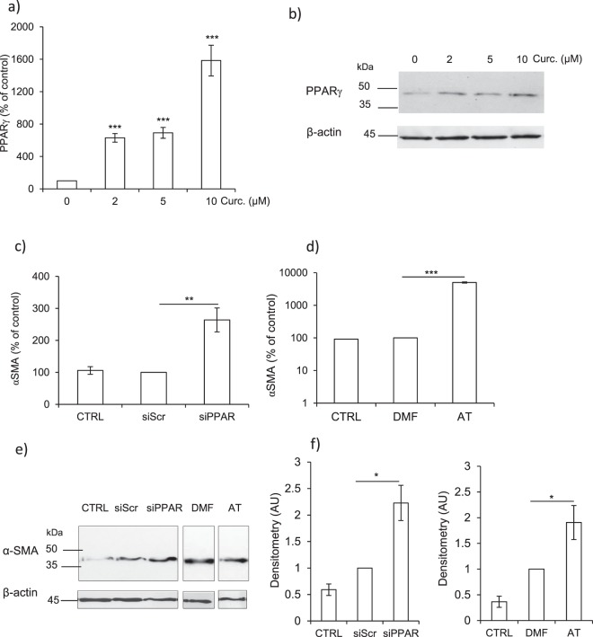
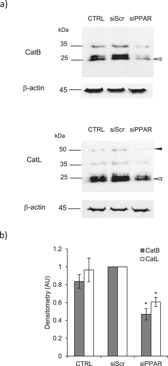
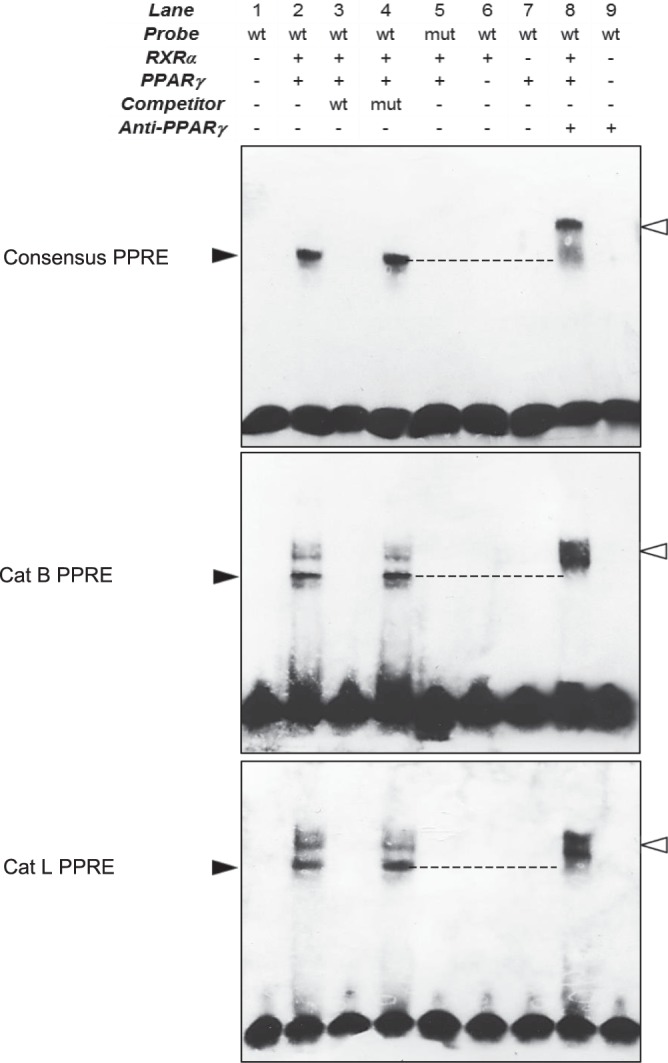
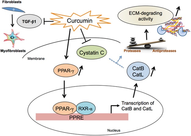
Pulmonary fibrosis is a progressive disease characterized by a widespread accumulation of myofibroblasts and extracellular matrix components. Growing evidences support that cysteine cathepsins, embracing cathepsin B (CatB) that affects TGF-β1-driven Smad pathway, along with their extracellular inhibitor cystatin C, participate in myofibrogenesis. Here we established that curcumin, a potent antifibrotic drug used in traditional Asian medicine, impaired the expression of both α-smooth muscle actin and mature TGF-β1 and inhibited the differentiation of human lung fibroblasts (CCD-19Lu cells). Curcumin induced a compelling upregulation of CatB and CatL. Conversely cystatin C was downregulated, which allowed the recovery of the peptidase activity of secreted cathepsins and the restoration of the proteolytic balance. Consistently, the amount of both insoluble and soluble type I collagen decreased, reaching levels similar to those observed for undifferentiated fibroblasts. The signaling pathways activated by curcumin were further examined. Curcumin triggered the expression of nuclear peroxisome proliferator-activated receptor γ (PPARγ). Contrariwise PPARγ inhibition, either by an antagonist (2-chloro-5-nitro-N-4-pyridinyl-benzamide) or by RNA silencing, restored TGF-β1-driven differentiation of curcumin-treated CCD-19Lu cells. PPARγ response element (PPRE)-like sequences were identified in the promoter regions of both CatB and CatL. Finally, we established that the transcriptional induction of CatB and CatL depends on the binding of PPARγ to PPRE sequences as a PPARγ/Retinoid X Receptor-α heterodimer.


Conflict of interest statement

The authors declare no competing interests.

Figures

Figure 1
Figure 1
Effect of curcumin on α-SMA expression of human CCD-19Lu myofibroblasts. (a) Three days after induction of the differentiation of CDD-19Lu cells into myofibroblasts by TGF-β1 (5 ng/ml), curcumin (0–50 µM) was added for different time intervals (24–96 h). The cell viability was then determined by MTS assay. Results (average values) were normalized using untreated cells as control (100% of viability) (n = 3). (b) Three days after addition of TGF-β1, myofibroblasts were treated with curcumin (0–10 µM) for 48 h. Analysis of α-SMA expression was performed by quantitative real time PCR analysis of α-SMA transcripts. Data are expressed as percentage relative to untreated control. (c) In parallel, myofibroblasts layers were harvested and lysed. Samples were submitted to electrophoresis (12% SDS-PAGE, under reducing conditions) before the protein level of α-SMA was analyzed by western blot using a mouse α-SMA antibody. β-actin was used for load control. A representative sample of three independent experiments is shown. Full-length blots are presented in Supplementary Fig. 5. (d) Corresponding WB densitometric analysis of the expression level of α-SMA; normalized data correspond to the average of three independent experiments, using β-actin as control. (e) Cells were treated with curcumin for 48 h as reported above. The green staining corresponds to the immunolabeling of α-SMA. Nuclei were stained by DAPI (blue). Scale bar represents 100 µm.
Figure 2
Figure 2
Consequences of curcumin treatment on profibrotic markers of lung myofibroblasts. (a) After treatment of CCD-19Lu cells with curcumin (0–10 µM) for 48 h, TGF-β1 transcripts were analyzed by quantitative real time PCR. Normalized data are expressed as percentage relative to untreated control (n = 3). (b) Western blot analysis of TGF-β1 expression level. A representative sample of three independent experiments is shown. White arrows indicate precursor pro-region forms; black arrows indicate dimer forms of TGF-β1. β-actin was used for load control. Full-length blots are presented in Supplementary Fig. 5. (c) Transcriptional analysis of Col1a1 (collagen type I, alpha-1; white bar) and Col1a2 (collagen type I, alpha-2; grey bar) mRNA by quantitative real time PCR analysis. Normalized data are expressed as percentage relative to untreated control (n = 3). (d) Dosage of soluble collagen in culture media (Sircol assay) (n = 3). (e) Dosage of insoluble collagen in cell lysates (Sircol assay) (n = 3).
Figure 3
Figure 3
Expression level of cathepsins B and L in CCD-19Lu myofibroblasts treated by curcumin. Three days after induction of the differentiation of CCD-19l-Lu cells by TGF-β1 (5 ng/ml), curcumin (0–10 µM) was added for 48 h. (a) Quantitative real time PCR analysis of CatB and CatL. mRNA levels are normalized and expressed as percentage relative to untreated control (n = 3). (b) The related peptidase activity of secreted cysteine cathepsins was measured using Z-Phe-Arg-AMC (50 µM) as substrate. Results (corresponding to the release of fluorescent AMC, reported as arbitrary unit) are normalized and expressed as percentage relative to control in the absence of curcumin treatment (n = 3). Active site labeling of extracellular cathepsins by Biotinyl-(PEG)2-Ahx-LVG-DMK. Culture media of CCD-19Lu cells were incubated for 1 h with the activity-based probe (10 µM) at 30 °C according to. Samples were subjected to electrophoresis on 12% SDS-PAGE under reducing conditions, electrotransferred to a nitrocellulose membrane, then blocked with 3% BSA in PBS-T. After incubation with an extravidin-peroxydase conjugate (1:3000, Sigma Aldrich) 2 h at room temperature, active cathepsins (lanes: 0, 2, 5, and 10 µM curcumin) were stained by chimiluminescence (ECL Plus Western Blotting Detection system). Full-length blots are presented in Supplementary Fig. 5. (c) Two days after addition of curcumin, myofibroblasts layers were lysed, and the expression of intracellular CatB and CatL was analyzed by western blotting. A representative sample is shown (n = 3). White arrows indicate mature forms; black arrows correspond to pro-CatB and pro-CatL. β-actin was used for load control. Full-length blots are presented in Supplementary Fig. 5. (d) Densitometric analysis of the protein level of intracellular mature CatB and CatL (normalized data relative to control without curcumin, n = 3). (e) Two days after treatment with curcumin, the protein level of extracellular CatB and CatL was analyzed by WB. A representative sample is shown (n = 3, white arrows, mature proteases). Full-length blots are presented in Supplementary Fig. 5. (f) Corresponding densitometric analysis of extracellular mature CatB and CatL. Normalized data relative to control without curcumin (n = 3).
Figure 4
Figure 4
Effect of curcumin on the expression of endogenous inhibitors of cysteine cathepsins. CCD-19Lu myofibroblasts were treated with curcumin (0–10 µM) for 48 h as previously reported. (a) Quantitative real time PCR analysis of cystatin C and stefin B. Normalized data are expressed as percentage relative to untreated control (n = 3). (b) Cystatin C ELISA. After retrieval of CCD-19Lu supernatants, the concentration of secreted cystatin C was measured by sandwich ELISA (DuoSet kit, R&D Systems) (n = 3). (c) Culture media were concentrated (x10), submitted to a 12% SDS-PAGE electrophoresis under reducing conditions and the protein level of extracellular cystatin C was analyzed by immunoblotting. Alternatively, cell lysates were prepared and intracellular stefin B expression examined in the same way by WB. A representative sample is shown (n = 3) and β-actin was used for load control. Full-length blots are presented in Supplementary Fig. 5. (d) Corresponding densitometric analysis (normalized data relative to control without curcumin, n = 3).
Figure 5
Figure 5
Overexpression of PPARγ in curcumin-treated myofibroblasts and consequences of PPARγ inhibition on the expression of α-SMA. CCD-19Lu myofibroblasts were treated with curcumin (0–10 µM) for 48 h as described earlier. (a) Quantitative real time PCR analysis of PPARγ. The data are expressed as percentage relative to untreated control (n = 3). (b) Myofibroblasts layers were harvested and lysed, and the expression of PPARγ analyzed by western blot. A representative sample of three independent experiments is shown and β-actin was used for load control. Full-length blots are presented in Supplementary Fig. 5. (c) Silencing of PPARγ by siRNA: six hours before addition of curcumin (10 µM), myofibroblasts were transfected with siRNA directed against PPARγ. α-SMA expression was analysed by qRT-PCR (CTRL, control, i.e. curcumin, no siRNA; siScr, control (scrambled) siRNA; siPPAR, siRNA directed against PPARγ; n = 3). (d) Pharmacological inhibition of PPARγ: six hours before curcumin treatment (10 µM), the pharmacological high-affinity PPARγ antagonist T0070907 (2-chloro-5-nitro-N-4-pyridinyl-benzamide, also called AT) was added to the culture medium. α-SMA expression was analyzed by qRT-PCR (CTRL, control, i.e. curcumin; DMF, vehicle; AT, cells treated with T0070907). Data are expressed as percentage relative to control (n = 3). (e) WB analysis of α-SMA after silencing of PPARγ and chemical inhibition of PPARγ. A representative sample is shown (n = 3) and β-actin was used for load control. Full-length blots are presented in Supplementary Fig. 5. (f) Respective densitometric analysis of WB (normalized data relative to control, n = 3).
Figure 6
Figure 6
Consequences of PPARγ inhibition on the expression of cathepsins B and L. CCD-19Lu myofibroblasts were transfected with siRNA directed against PPARγ (as described above) 6 h before curcumin treatment. Two days after, the expression of cathepsins was examined. (a) Immunoblotting analysis of intracellular CatB and CatL. White arrows indicate mature forms and black arrows indicate proforms of cathepsins. A representative sample is shown (n = 3) and β-actin was used for load control. Full-length blots are presented in Supplementary Fig. 5. (b) Densitometric analysis of CatB and CatL expression (Normalized Data, n = 3).
Figure 7
Figure 7
Binding of PPARγ to PPARγ response element-like (PPRE) sequences located in cathepsin B and L promoters. Electrophoretic mobility shift assays were performed using the end-labeled oligonucleotides representing the wild-type (wt) consensus PPRE, the human putative CatB PPRE-like sequences, the human putative CatL PPRE-like sequences (lanes 1, 2, 6 & 7) and mutated (mut) versions of these three sequences (lane 5) in the presence of human recombinant PPARγ and RXRα. Molar excess 200-fold (lanes 3 & 4) of unlabeled oligonucleotides was used for competition analysis. Supershift assays were performed using an anti-human PPARγ (lanes 8 & 9). Black arrows indicate specific gel shifts and white arrows indicate the supershifted bands (a representative sample is shown, n = 3). Full-length blots are presented in Supplementary Fig. 5.
Figure 8
Figure 8
Anti-fibrotic properties of curcumin are associated with PPARγ-driven upregulation of cathepsins B and L (a synthetic drawing). Curcumin inhibits the differentiation of lung CCD-19Lu fibroblasts (as confirmed by decreased amounts of both insoluble and soluble type I collagen, as well the impairment of expression levels of α-SMA, a profibrotic biomarker and endogenous TGF-β1). Moreover, curcumin triggers PPARγ that in turn may bind to PPARγ response element-like sequences, which are located in the promoter regions of CatB and CatL, and drives the transcription and expression of the two proteases. Otherwise curcumin down-regulates the expression level of cystatin C, the most potent circulating inhibitor of secreted cathepsins. Taken together this could allow the recovery of the proteolytic activity of secreted cathepsins and the restoration of the “cathepsins/cystatin C” balance, thus promoting ECM-degrading properties of cathepsins.

References

    1. Plantier, L. et al. Physiology of the lung in idiopathic pulmonary fibrosis. Eur Respir Rev27 (2018). - PMC - PubMed
    1. Noble PW, Barkauskas CE, Jiang D. Pulmonary fibrosis: patterns and perpetrators. J. Clin. Invest. 2012;122:2756–2762. doi: 10.1172/JCI60323. - DOI - PMC - PubMed
    1. Fernandez IE, Eickelberg O. New cellular and molecular mechanisms of lung injury and fibrosis in idiopathic pulmonary fibrosis. Lancet. 2012;380:680–688. doi: 10.1016/S0140-6736(12)61144-1. - DOI - PubMed
    1. Perez A, Rogers RM, Dauber JH. The prognosis of idiopathic pulmonary fibrosis. Am. J. Respir. Cell Mol. Biol. 2003;29:S19–26. doi: 10.1165/rcmb.2002-0254OC. - DOI - PubMed
    1. Sime PJ, Xing Z, Graham FL, Csaky KG, Gauldie J. Adenovector-mediated gene transfer of active transforming growth factor-beta1 induces prolonged severe fibrosis in rat lung. J. Clin. Invest. 1997;100:768–776. doi: 10.1172/JCI119590. - DOI - PMC - PubMed

Publication types

MeSH terms