. 2019 Jan 24;9(1):568.
doi: 10.1038/s41598-018-37071-y.

Fibrogenic Gene Expression in Hepatic Stellate Cells Induced by HCV and HIV Replication in a Three Cell Co-Culture Model System

Affiliations

Fibrogenic Gene Expression in Hepatic Stellate Cells Induced by HCV and HIV Replication in a Three Cell Co-Culture Model System

Abdellah Akil et al. Sci Rep. .

Abstract

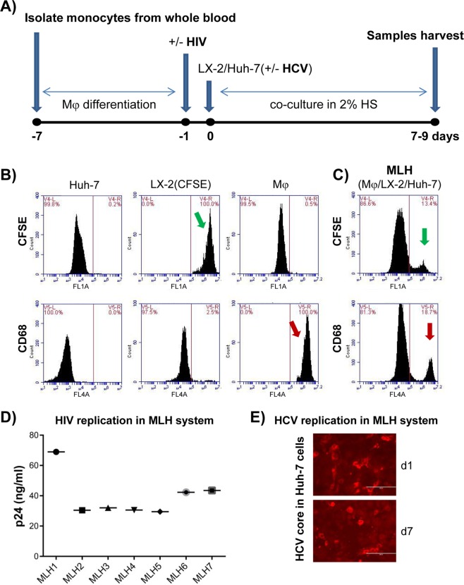
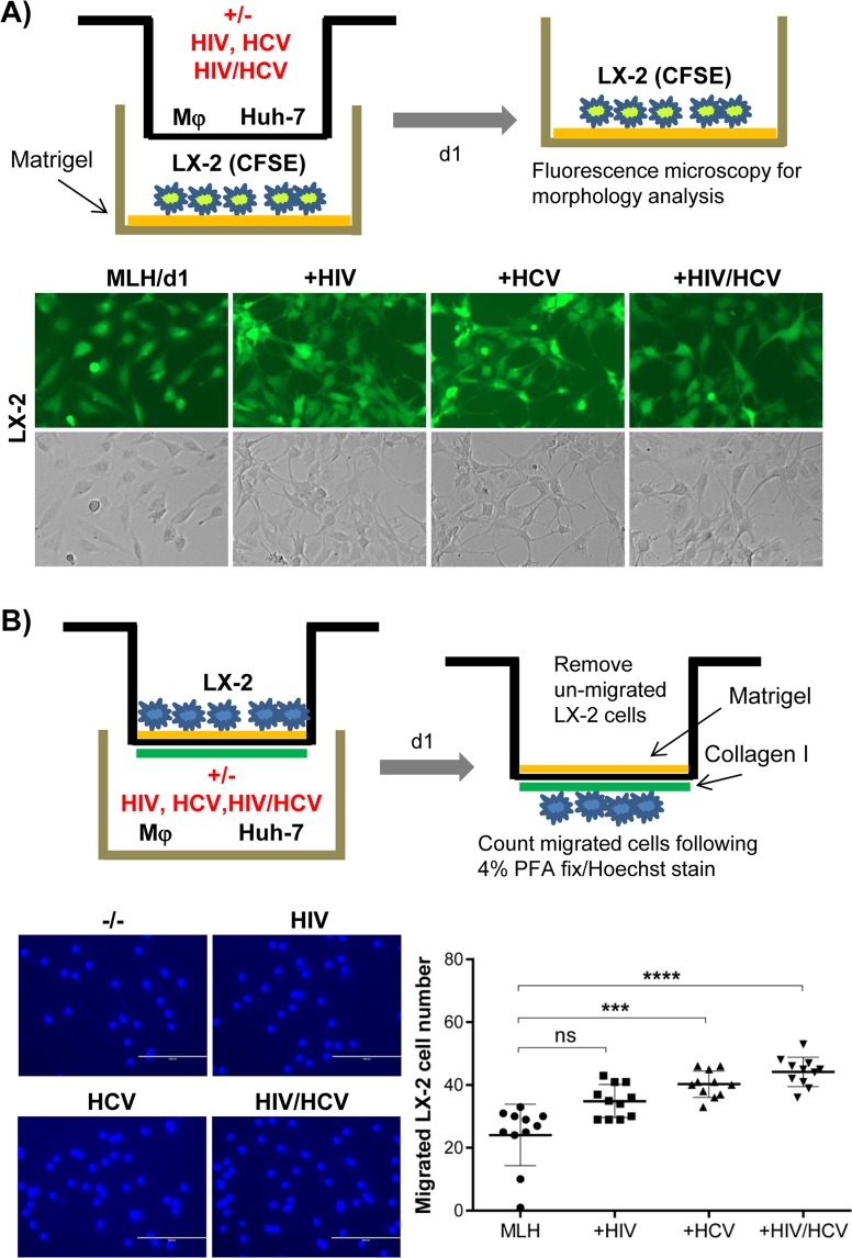
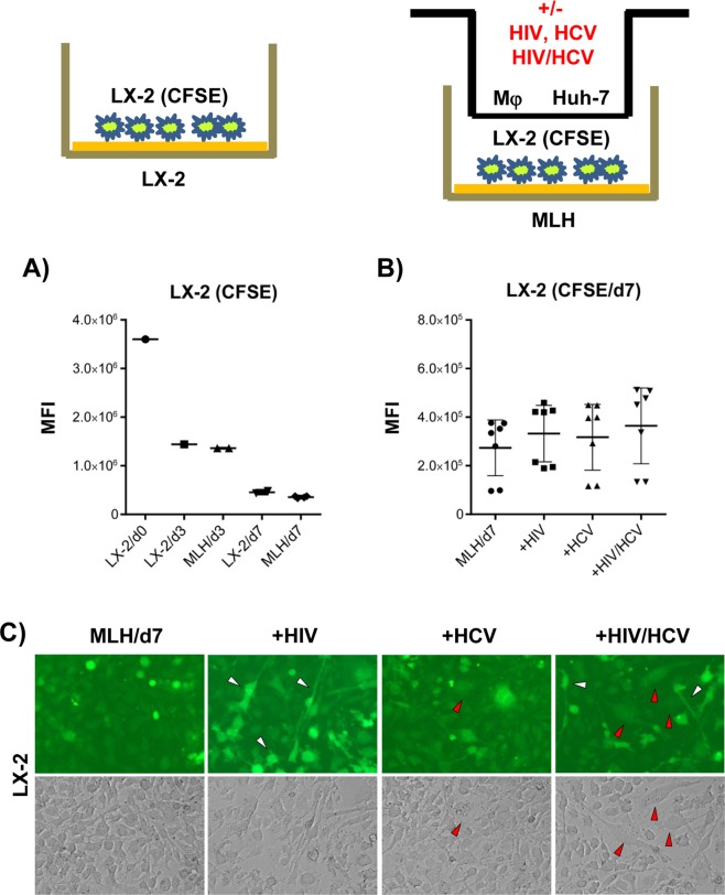
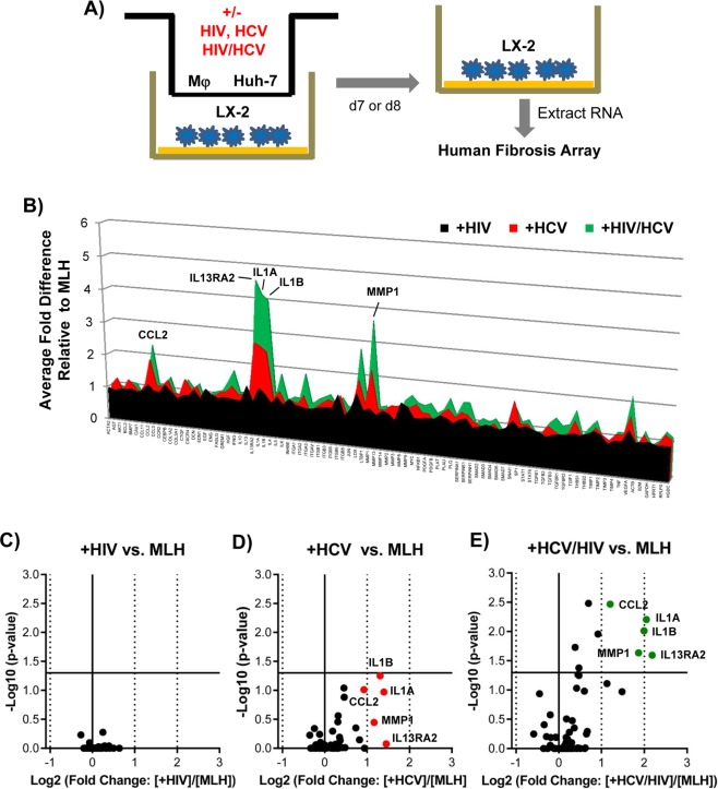
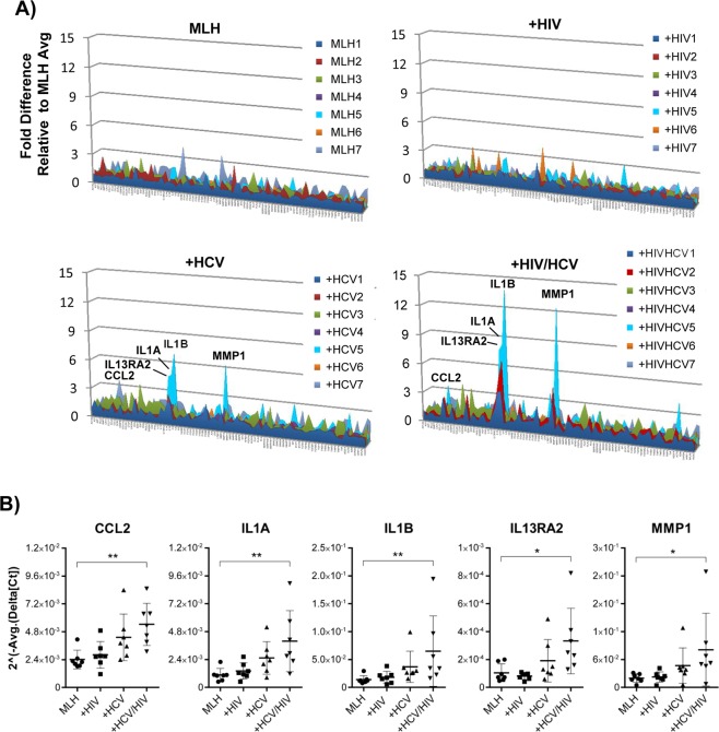
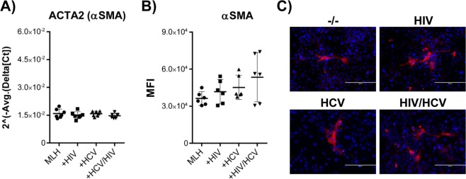
Retrospective studies indicate that co-infection of hepatitis C virus (HCV) and human immunodeficiency virus (HIV) accelerates hepatic fibrosis progression. We have developed a co-culture system (MLH) comprising primary macrophages, hepatic stellate cells (HSC, LX-2), and hepatocytes (Huh-7), permissive for active replication of HCV and HIV, and assessed the effect of these viral infections on the phenotypic changes and fibrogenic gene expression in LX-2 cells. We detected distinct morphological changes in LX-2 cells within 24 hr post-infection with HCV, HIV or HCV/HIV in MLH co-cultures, with migration enhancement phenotypes. Human fibrosis microarrays conducted using LX-2 cell RNA derived from MLH co-culture conditions, with or without HCV and HIV infection, revealed novel insights regarding the roles of these viral infections on fibrogenic gene expression in LX-2 cells. We found that HIV mono-infection in MLH co-culture had no impact on fibrogenic gene expression in LX-2 cells. HCV infection of MLH co-culture resulted in upregulation (>1.9x) of five fibrogenic genes including CCL2, IL1A, IL1B, IL13RA2 and MMP1. These genes were upregulated by HCV/HIV co-infection but in a greater magnitude. Conclusion: Our results indicate that HIV-infected macrophages accelerate hepatic fibrosis during HCV/HIV co-infection by amplifying the expression of HCV-dependent fibrogenic genes in HSC.


Conflict of interest statement

The authors declare no competing interests.

Figures

Figure 1
Figure 1
Development of three cell co-culture system (MLH) permissive for HCV and HIV replication consisting of macrophages (Mφ), hepatic stellate cells (HSC, LX-2) and hepatocytes (Huh-7). (A) Schematic of co-culture procedure. ‘HS’ denotes for human serum. (B) Huh-7, CFSE-labeled LX-2 and Alexa®647-CD68-labeled Mφ mono-cultures were subjected to FACS analysis. (C) FACS analysis following the MLH co-culture for 9 days. Green and red arrow indicate the detection of CFSE-labeled LX-2 and CD68-labeled Mφ at the end of co-culture. Majority of unlabeled cells belong to Huh-7 cells. (D) Replication of HIV under MLH co-culture for 7 to 8 days with Mφ derived from seven healthy volunteers detected by HIV p24 antigen Elisa assay. (E) Replication of HCV under MLH co-culture condition detected by using HCV core antigen staining.
Figure 2
Figure 2
HCV or HIV replication in MLH co-culture changed the morphological and invasive phenotypes of LX-2 cells. (A) Schematic of trans-well system separating LX-2 cells from Huh-7 and Mφ (top). Morphological characteristics of CFSE-labeled LX2 cells following MLH co-culture with or without HIV and HCV for 24 hr (bottom). (B) Schematic of LX2 cell invasion assay (top). Hoechst stained LX-2 cells detected at the bottom of trans-well insert following their migration (bottom left). The numbers of migrated LX-2 cells in three independent MLH co-cultures (bottom right). Asterisks indicate statistically significant difference measured by one-way ANOVA: ****p < 0.00005; ***p < 0.0005; ns, not significant.
Figure 3
Figure 3
Mixed morphological changes in LX-2 cells induced by HCV/HIV co-infection of MLH co-cultures. (A) Proliferation of CFSE-labeled LX-2 cells during mono-culture and MLH co-culture during 7 day culture period determined by using flow cytometry. Day 0 (d0) indicates the time for initiating LX-2(CFSE) culture individually or under the condition of MLH co-culture, and the CFSE levels in LX2/d0 and MLH/d0 (data not shown) are same. (B) Effects of HCV and/or HIV replication in MLH co-culture on CFSE-labeled LX-2 cell proliferation determined by using FACS at day 7 of MLH co-culture. Results are from three independent experiments. (C) Morphology of LX-2 cells under MLH co-culture observed at day 7 by using a fluorescence and phase-contrast microscope. (C) LX-2 cell morphology under MLH co-culture at day 7 detected using a fluorescent and phase-contrast microscope.
Figure 4
Figure 4
HCV/HIV co-infection augmented the induction of specific fibrogenic genes upregulated by HCV in MLH co-culture system. (A) Schematic of human fibrosis micro array by using LX-2 cell RNA isolated following MLH-co-culture with or without HCV and HIV. (B) Relative gene expression fold changes in LX-2 cells detected in the microarray analysis between non-infected and HIV-, HCV- and HCV/HIV-infected MLH co-cultures, respectively. A volcano plots showing the deregulated genes in LX-2 cells following (C) HIV, (D) HCV or (E) HCV/HIV infection of MLH co-culture. Genes up-regulated more than 1.9 folds by HCV and HCV/HIV infection are shown in red and green dots, respectively.
Figure 5
Figure 5
Primary Mφ in MLH co-cultures regulated the magnitude of HCV- or HCV/HIV-dependent fibrogenic gene induction in LX-2 cells. (A) Gene transcript levels in LX-2 cells from MLH co-cultures uninfected or infected with HCV and HIV, normalized to average values of individual genes from uninfected MLH co-cultures. (B) Individual values of normalized gene expression levels of five fibrogenic genes whose expression were upregulated more than 1.9 fold on average by HCV infection following 7 independent MLH co-cultures. Asterisks indicate statistically significant difference measured by one-way ANOVA: **p < 0.005; *p < 0.05.
Figure 6
Figure 6
Effect of HCV and/or HIV infection in MLH on αSMA expression in LX2 cells. (A) Normalized gene expression levels of αSMA gene in LX2 cells from seven different MLH co-cultures in the presence or absence of HCV and HIV. (B) FACS analysis of αSMA protein expression profile in LX-2 cells on day 7 of three independent MLH co-cultures with or without HCV and HIV infection. (C) Immunofluorescence analysis of αSMA in LX2 cells on day 7 of MLH co-cultures in the presence or absence of HCV and HIV.

References

    1. Bataller R, Brenner DA. Liver fibrosis. J Clin Invest. 2005;115:209–218. doi: 10.1172/JCI24282. - DOI - PMC - PubMed
    1. Hui AY, Friedman SL. Molecular basis of hepatic fibrosis. Expert Rev Mol Med. 2003;5:1–23. doi: 10.1017/S1462399403005684. - DOI - PubMed
    1. Friedman SL. Mechanisms of hepatic fibrogenesis. Gastroenterology. 2008;134:1655–1669. doi: 10.1053/j.gastro.2008.03.003. - DOI - PMC - PubMed
    1. Lee YA, Wallace MC, Friedman SL. Pathobiology of liver fibrosis: a translational success story. Gut. 2015;64:830–841. doi: 10.1136/gutjnl-2014-306842. - DOI - PMC - PubMed
    1. Seki E, Schwabe RF. Hepatic inflammation and fibrosis: functional links and key pathways. Hepatology. 2015;61:1066–1079. doi: 10.1002/hep.27332. - DOI - PMC - PubMed

Publication types

MeSH terms