. 2019 Jan 24;9(1):630.
doi: 10.1038/s41598-018-37198-y.

Liver Derived FGF21 Maintains Core Body Temperature During Acute Cold Exposure

Affiliations

Liver Derived FGF21 Maintains Core Body Temperature During Acute Cold Exposure

Magdalene Ameka et al. Sci Rep. .

Abstract

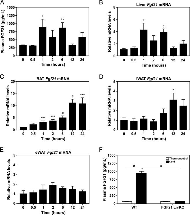
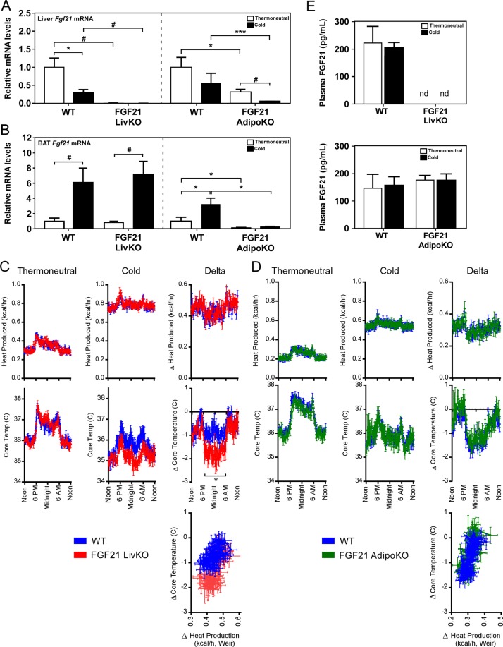
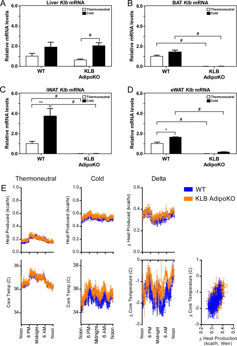
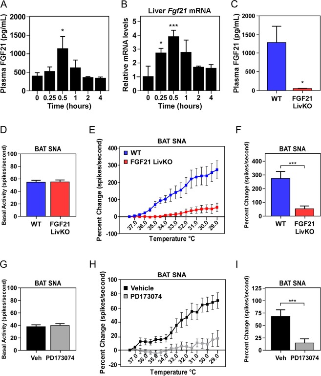
Fibroblast Growth Factor 21 (FGF21) elicits an array of metabolic effects. However, the physiological role of FGF21 during thermal challenges is not clear. In this study, we assessed the tissue source of FGF21 and its site of action to regulate core body temperature in response to cold. Using mice lacking FGF21 specifically in the liver (FGF21 LivKO) or adipose tissues (FGF21 AdipoKO), we performed a series of cold exposure studies to examine the tissue specific induction of FGF21 in response to cold. We also examined the physiological site of FGF21 action during cold exposure by impairing FGF21 signaling to adipose tissues or the central nervous system (CNS) using genetic ablation of the FGF21 co-receptor β-klotho in adipose tissues (KLB AdipoKO) or pharmacological blockage of FGF21 signaling. We found that only liver-derived FGF21 enters circulation during acute cold exposure and is critical for thermoregulation. While FGF21 signaling directly to adipose tissues during cold is dispensable for thermoregulation, central FGF21 signaling is necessary for maximal sympathetic drive to brown adipose tissue to maintain thermoregulation during cold. These data demonstrate a previously unrecognized role for FGF21 in the maintenance of body temperature in response to cold.


Conflict of interest statement

The authors declare no competing interests.

Figures

Figure 1
Figure 1
Acute cold exposure increases circulating levels of FGF21. (A) Plasma FGF21 levels in 12 week old C57Bl/6J male mice cold exposed for the indicated amount of time (n = 7/group). (B-E) Fgf21 mRNA levels in (B) liver, (C) BAT, (D) iWAT and (E) eWAT from mice in (A). (F) Plasma FGF21 levels in 11–13 week old wild type (WT) and FGF21 LivKO male mice cold exposed for 1 hour (n = 5–6/group). Values are mean ± SEM; *P ≤ 0.05; **P ≤ 0.01; ***P ≤ 0.005; and #P ≤ 0.001 compared to time 0 for (A–E) and relative to WT for (F).
Figure 2
Figure 2
Hepatic FGF21 production is critical to maintain core body temperature during cold exposure. (A) Liver and (B) brown adipose tissue (BAT) Fgf21 mRNA levels in 11–13 week old wild type (WT), FGF21 LivKO and FGF21 AdipoKO male mice housed at thermoneutrality or following 3 days of cold exposure. (C,D) Core body temperature (°C) and heat production (kcal/h) during thermoneutral and cold phases of 11–13 week old male FGF21 LivKO (C) and FGF21 AdipoKO (D) mice relative to WT controls (n = 6–8/group). Heat production by hour is estimated by the modified Weir equation. Core body temperature (°C) is also plotted against change in heat production (kcal/h). Change in core body temperature and heat production relative to time of day is also presented for FGF21 LivKO (C) and FGF21 AdipoKO (D) mice compared to WT controls (n = 6–8/group). For C, Genotype: P = 0.0866, Interaction between genotype and time of day: P = 0.0361 by 2-way RM ANOVA. (E) Plasma FGF21 levels from FGF21 LivKO and FGF21 AdipoKO mice shown in (C,D). Values are mean ± SEM; *P ≤ 0.05; **P ≤ 0.01; ***P ≤ 0.005; and #P ≤ 0.001 compared to wild type mice; nd = not detected.
Figure 3
Figure 3
Loss of FGF21 signaling to adipose tissues does not affect core body temperature during cold exposure. (A) Liver, (B) BAT, (C) iWAT and (D) eWAT Klb mRNA levels in 11–13 week old male wild type (WT) and KLB AdipoKO mice housed at thermoneutrality or following 3 days of cold exposure (n = 6–7/group). (E) Core body temperature (°C) and heat production (kcal/h) during thermoneutral and cold phases of 11–13 week old male KLB AdipoKO mice relative to WT controls (n = 6–7/group). Heat production by hour is estimated by the modified Weir equation. Core body temperature (°C) is also plotted against change in heat production (kcal/h). Change in core body temperature and heat production relative to time of day is also presented for KLB AdipoKO mice compared to WT controls (n = 6–7/group). Values are mean ± SEM; *P ≤ 0.05; **P ≤ 0.01; ***P ≤ 0.005; and #P ≤ 0.001 compared to wild type mice.
Figure 4
Figure 4
Liver-derived FGF21 regulates sympathetic nerve activity to BAT during cold exposure. (A) Plasma FGF21 levels and (B) hepatic Fgf21 mRNA levels in 12–14 week old male wild type (WT) C57Bl/6J mice administered norepinephrine (1 mg/kg) for the indicated time (n = 8/group). (C) Plasma FGF21 levels in WT and FGF21 LivKO mice 30 min post-norepinephrine injection (n = 5–9/group). (D) Basal SNA to brown adipose tissue (BAT) in 12–14 week old male WT and FGF21 LivKO mice at ambient temperature and (E) during incremental cooling (n = 6–7/group). (F) Final percent change in BAT SNA in wild type and FGF21 LivKO mice at the final cooling point of 29 °C. (G) Basal SNA to BAT in 13 week old male WT mice administered ICV vehicle or PD173074 (25 µg) or (H) during incremental cooling (n = 10–11/group). (I) Final percent change in BAT SNA at 29 °C in wild type mice administered vehicle or PD173074. Values are mean ± SEM; *P ≤ 0.05; **P ≤ 0.01; and ***P ≤ 0.005 compared to time 0 for (A,B) and relative to wild type mice (C–I).

Similar articles

Cited by

References

    1. Morrison, S. F. Central control of body temperature. F1000Res5, 10.12688/f1000research.7958.1 (2016). - PMC - PubMed
    1. Townsend KL, Tseng YH. Brown fat fuel utilization and thermogenesis. Trends in endocrinology and metabolism: TEM. 2014;25:168–177. doi: 10.1016/j.tem.2013.12.004. - DOI - PMC - PubMed
    1. Morrison SF, Madden CJ. Central nervous system regulation of brown adipose tissue. Compr Physiol. 2014;4:1677–1713. doi: 10.1002/cphy.c140013. - DOI - PMC - PubMed
    1. Cannon B, Nedergaard J. Brown adipose tissue: function and physiological significance. Physiol Rev. 2004;84:277–359. doi: 10.1152/physrev.00015.2003. - DOI - PubMed
    1. Oelkrug R, Polymeropoulos ET, Jastroch M. Brown adipose tissue: physiological function and evolutionary significance. J Comp Physiol B. 2015;185:587–606. doi: 10.1007/s00360-015-0907-7. - DOI - PubMed

Publication types

Substances