

Characterization of a novel species of adenovirus from Japanese microbat and role of CXADR as its entry factor
- PMID: 30679679
- PMCID: PMC6345744
- DOI: 10.1038/s41598-018-37224-z
Characterization of a novel species of adenovirus from Japanese microbat and role of CXADR as its entry factor
Abstract
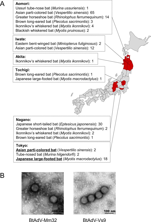
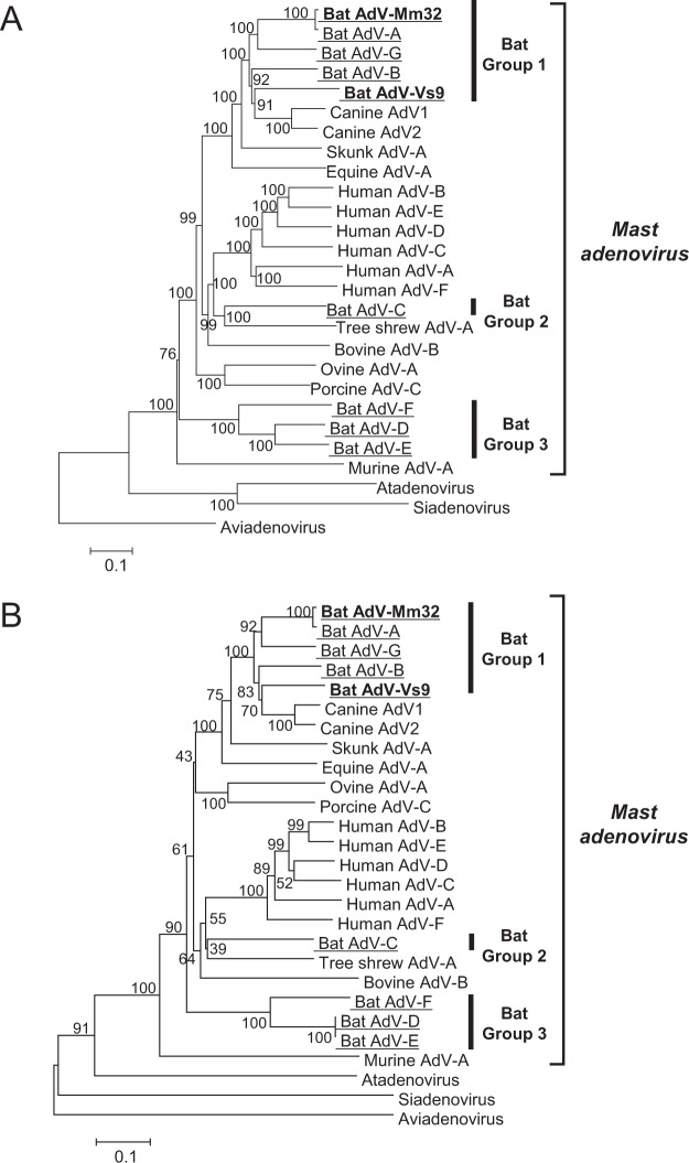
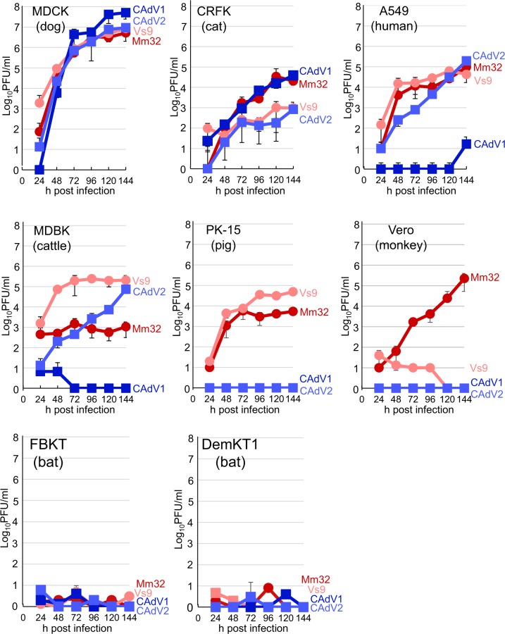
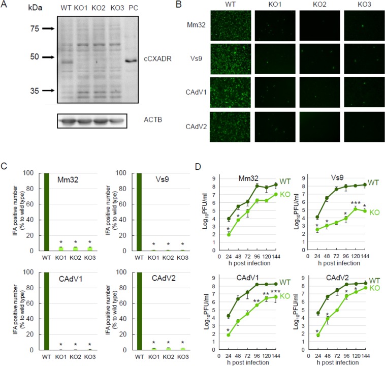
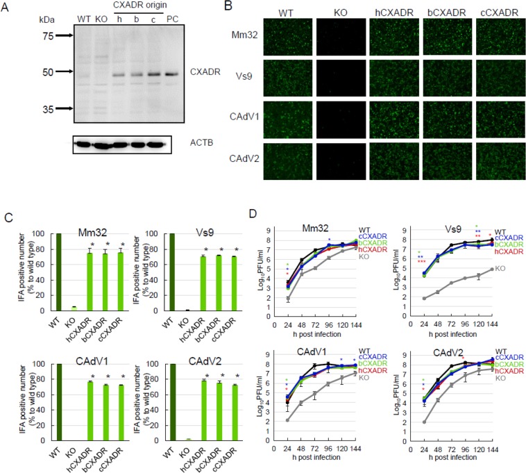
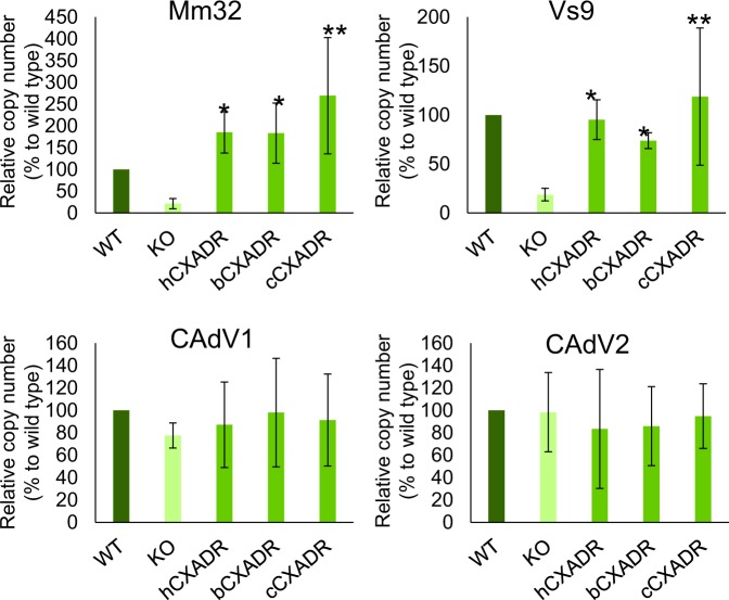
Recently, bat adenoviruses (BtAdVs) of genus Mastadenovirus have been isolated from various bat species, some of them displaying a wide host range in cell culture. In this study, we isolated two BtAdVs from Japanese wild microbats. While one isolate was classified as Bat mastadenovirus A, the other was phylogenetically independent of other BtAdVs. It was rather related to, but serologically different from, canine adenoviruses. We propose that the latter, isolated from Asian parti-colored bat, should be assigned to a novel species of Bat mastadenovirus. Both isolates replicated in various mammalian cell lines, implying their wide cell tropism. To gain insight into cell tropism of these BtAdVs, we investigated the coxsackievirus and adenovirus receptor (CXADR) for virus entry to the cells. We prepared CXADR-knockout canine kidney cells and found that replication of BtAdVs was significantly hampered in these cells. For confirmation, their replication in canine CXADR-addback cells was rescued to the levels with the original cells. We also found that viral replication was corrected in human or bat CXADR-transduced cells to similar levels as in canine CXADR-addback cells. These results suggest that BtAdVs were able to use several mammalian-derived CXADRs as entry factors.
Conflict of interest statement
The authors declare no competing interests.
Figures
Similar articles
-
Evolution and Cryo-electron Microscopy Capsid Structure of a North American Bat Adenovirus and Its Relationship to Other Mastadenoviruses.J Virol. 2017 Jan 3;91(2):e01504-16. doi: 10.1128/JVI.01504-16. Print 2017 Jan 15. J Virol. 2017. PMID: 27807242 Free PMC article.
-
Characterization of a novel adenovirus isolated from a skunk.Virology. 2015 Nov;485:16-24. doi: 10.1016/j.virol.2015.06.026. Epub 2015 Jul 17. Virology. 2015. PMID: 26189043
-
Isolation of a novel adenovirus from Rousettus leschenaultii bats from India.Intervirology. 2012;55(6):488-90. doi: 10.1159/000337026. Epub 2012 May 3. Intervirology. 2012. PMID: 22572722 Free PMC article.
-
Murine adenoviruses: tools for studying adenovirus pathogenesis in a natural host.FEBS Lett. 2019 Dec;593(24):3649-3659. doi: 10.1002/1873-3468.13699. Epub 2019 Dec 6. FEBS Lett. 2019. PMID: 31777948 Free PMC article. Review.
-
Coxsackievirus and Adenovirus Receptor (CXADR): Recent Findings and Its Role and Regulation in Spermatogenesis.Adv Exp Med Biol. 2021;1288:95-109. doi: 10.1007/978-3-030-77779-1_5. Adv Exp Med Biol. 2021. PMID: 34453733 Review.
Cited by
-
Dispersal history of Miniopterus fuliginosus bats and their associated viruses in east Asia.PLoS One. 2021 Jan 14;16(1):e0244006. doi: 10.1371/journal.pone.0244006. eCollection 2021. PLoS One. 2021. PMID: 33444317 Free PMC article.
-
Direct evidence of fiber-protein-directed hemagglutination by canine adenoviruses.Arch Virol. 2023 Feb 16;168(3):93. doi: 10.1007/s00705-023-05718-5. Arch Virol. 2023. PMID: 36795171 Free PMC article.
-
Complete genome sequence of a novel bat mastadenovirus C strain isolated from Rhinolophus cornutus in Japan.Arch Virol. 2022 Mar;167(3):979-982. doi: 10.1007/s00705-021-05357-8. Epub 2022 Feb 3. Arch Virol. 2022. PMID: 35112204 Free PMC article.
-
Serum proteomics reveals a tolerant immune phenotype across multiple pathogen taxa in wild vampire bats.Front Immunol. 2023 Dec 12;14:1281732. doi: 10.3389/fimmu.2023.1281732. eCollection 2023. Front Immunol. 2023. PMID: 38193073 Free PMC article.
-
Serological evidence of African pygmy hedgehog adenovirus 1 in exotic companion animals.J Vet Med Sci. 2025 Jul 11;87(7):862-867. doi: 10.1292/jvms.25-0164. Epub 2025 Jun 2. J Vet Med Sci. 2025. PMID: 40451840 Free PMC article.
References
Publication types
MeSH terms
Substances
LinkOut - more resources
Full Text Sources
Research Materials
