. 2019 Jan 24;9(1):600.
doi: 10.1038/s41598-018-37306-y.

The structure of the nucleoprotein of Influenza D shows that all Orthomyxoviridae nucleoproteins have a similar NPCORE, with or without a NPTAIL for nuclear transport

Affiliations

The structure of the nucleoprotein of Influenza D shows that all Orthomyxoviridae nucleoproteins have a similar NPCORE, with or without a NPTAIL for nuclear transport

Amélie Donchet et al. Sci Rep. .

Abstract

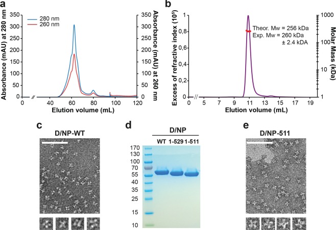
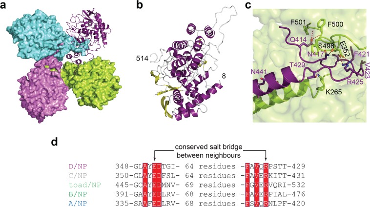
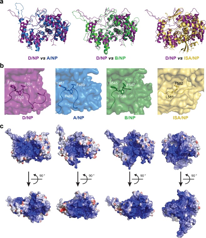
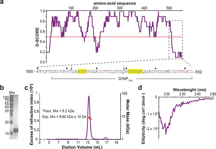
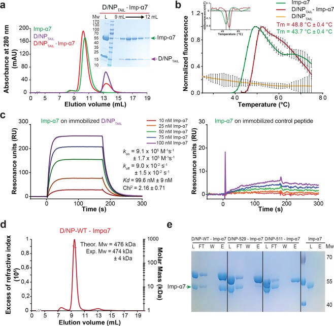
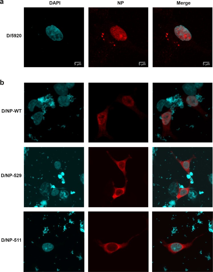
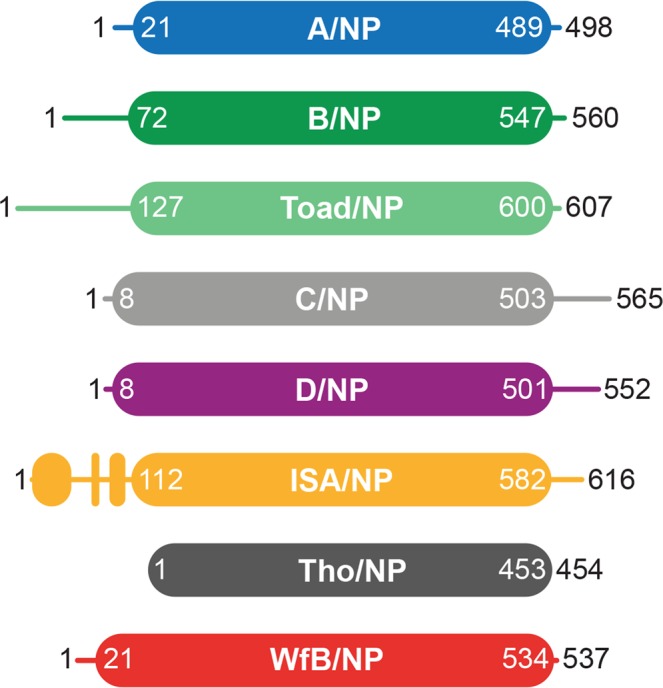
This paper focuses on the nucleoprotein (NP) of the newly identified member of the Orthomyxoviridae family, Influenza D virus. To date several X-ray structures of NP of Influenza A (A/NP) and B (B/NP) viruses and of infectious salmon anemia (ISA/NP) virus have been solved. Here we purified, characterized and solved the X-ray structure of the tetrameric D/NP at 2.4 Å resolution. The crystal structure of its core is similar to NP of other Influenza viruses. However, unlike A/NP and B/NP which possess a flexible amino-terminal tail containing nuclear localization signals (NLS) for their nuclear import, D/NP possesses a carboxy-terminal tail (D/NPTAIL). We show that D/NPTAIL harbors a bipartite NLS and designed C-terminal truncated mutants to demonstrate the role of D/NPTAIL for nuclear transport.


Conflict of interest statement

The authors declare no competing interests.

Figures

Figure 1
Figure 1
Purification and characterized of Influenza D nucleoprotein. (a) Size exclusion chromatography profile of wild-type D/NP. The sample was loaded on a HiloadTM 16/600 S200 column equilibrated with the running buffer 20 mM Tris-HCl pH 7.5, 300 mM NaCl and 5 mM β-mercaptoethanol. (b) SEC-MALLS-RI analysis of D/NP. SEC was performed with a SuperdexTM 200 increase 10/300 GL column equilibrated with 20 mM Tris-HCl pH 7.5, 150 mM NaCl and 5 mM β-ME. The panel shows the theoretical Mw and the measured Mw. (c) and (e) Electron microscopy images of the elution peak of D/NP and D/NP-511. Samples show different oligomeric states although most oligomers are tetramers. The scale bar corresponds to 100 nm. (d) Coomassie blue-stained SDS-PAGE (4–20% gradient polyacrylamide) showing the purified wild-type D/NP and the two C-terminal truncated mutants (D/NP-529 and D/NP-511).
Figure 2
Figure 2
Structure of Influenza D nucleoprotein. (a) Structure of the tetrameric D/NP, with 3 protomers shown in surface (respectively in green, pink and cyan) and the fourth in cartoon (deep purple). (b) Cartoon representation of one monomer of D/NP with the α-helixes in deep purple and β-strands in yellow. (c) Detail of the interactions between two protomers of the tetrameric D/NP as shown in (a). The conserved R425 of one protomer (shown as deep purple cartoon) stabilized the position of the oligomerization loop at the surface of the neighbouring protomer, through its conserved E352. (d) Sequence alignment of the salt bridges of the oligomerization-loop of one protomer to the NPCORE of the neighbour protomers. For the sequences see Table 1.
Figure 3
Figure 3
Comparison of D/NP with other segmented negative sRNA virus nucleoproteins. (a) The structure of one protomer of D/NP (deep purple) has been superimposed from left to right, with one protomer of A/NP (blue; PDB id: 2IQH), B/NP (forest; PDB id: 3TJ0) and ISA/NP (light orange; PDB id: 4EWC). The rmsd values are given in Table 1. (b) Anchoring of the C-terminus on NPCORE by a patch of 3 consecutive aromatic residues. The panel corresponds to a zoom of the superimposed structures shown in panel A with NPCORE represented in surface. (c) Electrostatic surface potentials of one protomer of D/NP, A/NP, B/NP and ISA/NP. The electrostatic surfaces were calculated from the crystal structures using DelPhi. The potential scales range from −10.0 kT/e (red) to 10.0 kT/e (blue).
Figure 4
Figure 4
Biophysical characterization of D/NPTAIL. (a) D-score (score for disorder as a function of residue) of D/NP with a zoom (below the graph) on the last 50 residues. The prediction is based on 22 predictor web servers and the D-score was calculated by adding the values for each residue and dividing by the number of used algorithms. We arbitrarily defined a threshold level at 0.50; residues with a D-score <0.50 were assigned as disordered. The yellow boxes on the sequence are to highlight the putative NLS motifs. The arrows indicate where the sequences were cut for making D/NP-529 and D/NP-511. (b) Coomassie blue-stained SDS-PAGE (Tris-Tricine; 15% polyacrylamide) of the purified D/NPTAIL. It migrates at a higher molecular weight (17 kDa approximately) than expected (8 kDa). (c) SEC-MALLS-RI analysis of D/NPTAIL loaded on S75 10/300 GL column. For this experiment, we have chosen to keep the His-tag encoded with the pETM11 plasmid, for an optimal detection of D/NPTAIL with UV. The experimental molecular weight is consistent with the expected mass. (d) Circular dichroïsm of D/NPTAIL. CD is a biophysical method based on the polarization of light, used for a fast determination of the secondary structures within the proteins in solution. α-Helical proteins show negative bands at 222 nm and 208 nm and a positive band at 193 nm, proteins with well-defined antiparallel β-sheets have negative bands at 218 nm and positive bands at 195 nm and disordered proteins have very low ellipticity above 210 nm and negative bands near 195 nm.
Figure 5
Figure 5
Interaction of D/NP and D/NPTAIL with importin-α7. (a) Size exclusion chromatography profile of a mixture between human importin-α7 and D/NPTAIL. The mixture (molar ratio 1 importin-α7 for 2 D/NPTAIL) was incubated 1 hour at room temperature and then loaded on a SuperdexTM75 10/300GL column equilibrated with the running buffer 20 mM Tris-HCl pH 7.5, 250 mM NaCl, 5 mM β-mercaptoethanol. (b) Thermal stability assay of importin-α7 in absence (green) or in presence (red) of D/NPTAIL using Thermofluor. In presence of D/NPTAIL, the melting temperature of importin-α7 is 5 °C higher. D/NPTAIL alone using Thermofluor did not give any denaturation signal (yellow curve). The upper insert corresponds to the derivative of the fluorescence signal for a precise measure of the melting temperature. (c) Affinity of importin-α7 for D/NPTAIL by measured by surface plasmon resonance (SPR). Biotinylated D/NPTAIL (left) and control peptide (right) were captured on a streptavidin-coated sensor chip surface before injections of several importin-α7 concentrations (10 nM in red, 25 nM in orange, 50 nM in green, 75 nM in blue and 100 nM in purple). The sensorgrams of the interaction between D/NPTAIL and importin-α7 were fitted under a Langmuir 1:1 binding model with mass-transfer (black line). (d) SEC-MALLS analysis of D/NP in complex with importin-α7. The mixture (molar ratio 1 D/NP for 1.2 importin-α7) was incubated 1 hour at room temperature and then loaded on a SuperdexTM 200 increase 10/300 GL. The experimental molecular weight is consistent with the expected mass of four importins-α7 bound per D/NP tetramer. (e) Pull-down assays of human importin-α7 by D/NP and the two C-terminal truncated mutants (D/NP-529 and D/NP-511). The his-tags are on D/NP. The mixtures (molar ratio 1 D/NP for 1.2 importin-α7) were incubated 1 hour and the experience was done as described in panel (a). The figure shows the coomassie blue-stained SDS-PAGE (12% polyacrylamide) with the Load, FlowThough, Wash and the second fractions (E2).
Figure 6
Figure 6
Nuclear transport of D/NP in HEK 293T cells. Microscopy pictures of HEK 293T cells with DAPI, NP and merged DAPI + NP staining (left, middle and right panels, respectively). (a) D/NP after infection of HEK 293T cells at 6 hours post infection with a moi of 5. (b) Cells were transfected with wt D/NP, D/NP-529 and D/NP-511. After 24 h cells were fixed, permeabilized and analyzed by indirect immunofluorescence. NP localization was observed using an in house rabbit hyperimmune NP-IDV serum and anti-rabbit IgG labeled Rhodamine RX (in red). Cells were mounted with DAPI-Vectashield and observed with a Leica Zeiss 710 (magnification: x63). In red: NP protein; in blue: nucleus. The bar in (A) represents 5 µm.
Figure 7
Figure 7
Schemas for nucleoproteins of Orthomyxoviruses. Schematic representation of the nucleoprotein based on the amino acid sequence identity and structure analysis of the protein from representative members of the Orthomyxoviridae family. The schema respects the size of the proteins. The protein accession numbers are the same as Table 1. The flexible tails are represented with simple lines whereas the folded parts and the cores are represented with filled boxes.

References

    1. Ferguson L, et al. Influenza D virus infection in Mississippi beef cattle. Virology. 2015;486:28–34. doi: 10.1016/j.virol.2015.08.030. - DOI - PMC - PubMed
    1. Hause BM, et al. Isolation of a novel swine influenza virus from Oklahoma in 2011 which is distantly related to human influenza C viruses. PLoS Pathog. 2013;9:e1003176. doi: 10.1371/journal.ppat.1003176. - DOI - PMC - PubMed
    1. Luo J, et al. Serological evidence for high prevalence of Influenza D Viruses in Cattle, Nebraska, United States, 2003-2004. Virology. 2017;501:88–91. doi: 10.1016/j.virol.2016.11.004. - DOI - PMC - PubMed
    1. Mitra N, Cernicchiaro N, Torres S, Li F, Hause BM. Metagenomic characterization of the virome associated with bovine respiratory disease in feedlot cattle identified novel viruses and suggests an etiologic role for influenza D virus. J Gen Virol. 2016;97:1771–1784. doi: 10.1099/jgv.0.000492. - DOI - PMC - PubMed
    1. Horimoto T, et al. Nationwide Distribution of Bovine Influenza D Virus Infection in Japan. PLoS One. 2016;11:e0163828. doi: 10.1371/journal.pone.0163828. - DOI - PMC - PubMed

Publication types