

Aberrant CEACAM19 expression is associated with metastatic phenotype in penile cancer
- PMID: 30679925
- PMCID: PMC6338120
- DOI: 10.2147/CMAR.S192385
Aberrant CEACAM19 expression is associated with metastatic phenotype in penile cancer
Abstract
Objective: A greater knowledge of the mechanisms of the pathogenesis of penile cancers may assist in the development of more tailored targeted therapy. Herein, we aimed to evaluate the expression of CEACAM19 in penile cancer and to explore its regulatory mechanisms.
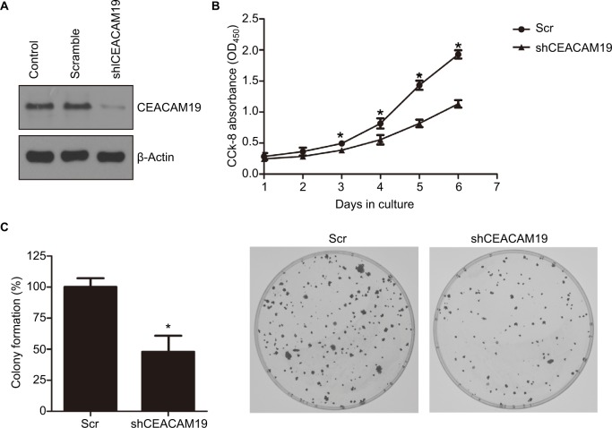
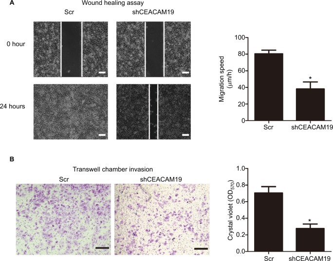
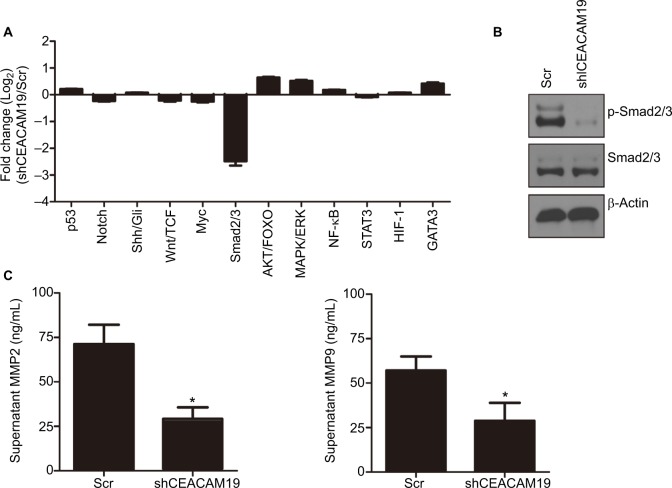
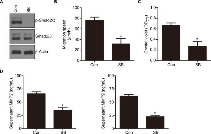
Material and methods: This retrospective study enrolled 64 penile cancer patients who underwent penectomy between 2011 and 2015. CEACAM19 expression in tissues was detected by immunohistochemistry, which was analyzed in association with clinicopathological parameters. Kaplan-Meier analysis was performed to evaluate the relationship between CEACAM19 expression and prognosis of patients with penile cancer. Cell Counting Kit-8 assay and clonogenic assay were used to evaluate the cell viability and tumorigenic potential of penile cancer cell line, respectively; wound healing assay and transwell invasion assay were conducted to evaluate the effect of CEACAM19 depletion on cell migration and invasion in penile cancer cells; CEACAM19 protein expression was analyzed by Western blotting. Culture supranatant matrix metalloproteinase 2/9 (MMP2/9) was detected by ELISA.
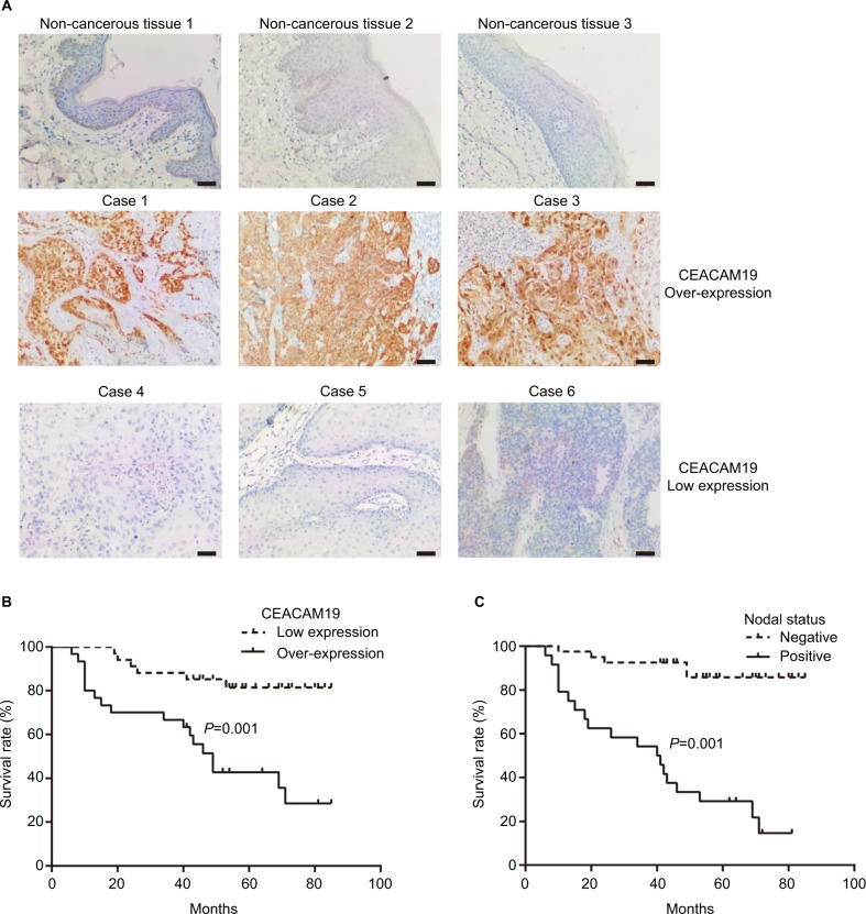
Results: CEACAM19 was differentially expressed in non-cancerous and penile cancer tissues. Over-expression of CEACAM19 was significantly associated with nodal and distant metastasis, and predicted unfavorable cancer-specific survival in penile cancer. Depletion of CEACAM19 expression suppressed cell proliferation, reduced colony formation, and attenuated cell migration and invasion in Penl1 cells. Furthermore, knockdown of CEACAM19 expression attenuated the levels of p-Smad2/3 and reduced secretion of MMP2/9 in Penl1 cells. The effects of CEACAM19 might result from its function in regulating the Smad2/3 activation, as inhibition on Smad2/3 activation suppressed cell migration and invasion and reduced MMP2/9 secretion in Penl1 cells.
Conclusion: Over-expression of CEACAM19 might serve as a potential prognostic biomarker for clinical management of penile cancer. Strategies targeting CEACAM19-regulated signaling pathways may have a therapeutic benefit in penile cancer.
Keywords: CEACAM19; metastasis; penile cancer; prognosis.
Conflict of interest statement
Disclosure The authors report no conflicts of interests in this work.
Figures
Similar articles
-
Serum CXCL13 Level is Associated with Tumor Progression and Unfavorable Prognosis in Penile Cancer.Onco Targets Ther. 2020 Aug 31;13:8757-8769. doi: 10.2147/OTT.S263980. eCollection 2020. Onco Targets Ther. 2020. PMID: 32943882 Free PMC article.
-
Preoperative plasma IGFBP2 is associated with nodal metastasis in patients with penile squamous cell carcinoma.Urol Oncol. 2019 Jul;37(7):452-461. doi: 10.1016/j.urolonc.2019.04.013. Epub 2019 Apr 30. Urol Oncol. 2019. PMID: 31053522
-
Knockdown of CEACAM19 suppresses human gastric cancer through inhibition of PI3K/Akt and NF-κB.Surg Oncol. 2018 Sep;27(3):495-502. doi: 10.1016/j.suronc.2018.05.003. Epub 2018 May 3. Surg Oncol. 2018. PMID: 30217308
-
Overexpression of ID1 promotes tumor progression in penile squamous cell carcinoma.Oncol Rep. 2019 Feb;41(2):1091-1100. doi: 10.3892/or.2018.6912. Epub 2018 Dec 6. Oncol Rep. 2019. PMID: 30535485
-
The expression of the CEACAM19 gene, a novel member of the CEA family, is associated with breast cancer progression.Int J Oncol. 2013 May;42(5):1770-7. doi: 10.3892/ijo.2013.1860. Epub 2013 Mar 21. Int J Oncol. 2013. PMID: 23525470
Cited by
-
Experimental Models for Studying HPV-Positive and HPV-Negative Penile Cancer: New Tools for An Old Disease.Cancers (Basel). 2021 Jan 26;13(3):460. doi: 10.3390/cancers13030460. Cancers (Basel). 2021. PMID: 33530343 Free PMC article. Review.
-
Serum CXCL13 Level is Associated with Tumor Progression and Unfavorable Prognosis in Penile Cancer.Onco Targets Ther. 2020 Aug 31;13:8757-8769. doi: 10.2147/OTT.S263980. eCollection 2020. Onco Targets Ther. 2020. PMID: 32943882 Free PMC article.
-
High serum CCL20 is associated with tumor progression in penile cancer.J Cancer. 2020 Sep 30;11(23):6812-6822. doi: 10.7150/jca.48939. eCollection 2020. J Cancer. 2020. PMID: 33123272 Free PMC article.
-
SHCBP1 regulates STAT3/c-Myc signaling activation to promote tumor progression in penile cancer.Am J Cancer Res. 2020 Oct 1;10(10):3138-3156. eCollection 2020. Am J Cancer Res. 2020. PMID: 33163262 Free PMC article.
References
-
- Paner GP, Stadler WM, Hansel DE, Montironi R, Lin DW, Amin MB. Updates in the eighth edition of the tumor-node-metastasis staging classification for urologic cancers. Eur Urol. 2018;73(4):560–569. - PubMed
-
- Ottenhof SR, Leone AR, Horenblas S, Spiess PE, Vegt E. Advancements in staging and imaging for penile cancer. Curr Opin Urol. 2017;27(6):612–620. - PubMed
-
- Ficarra V, Novara G, Boscolo-Berto R, Artibani W, Kattan MW. How accurate are present risk group assignment tools in penile cancer? World J Urol. 2009;27(2):155–160. - PubMed
LinkOut - more resources
Full Text Sources
Miscellaneous
