. 2019 Jan 22:16:3.
doi: 10.1186/s12014-019-9223-7. eCollection 2019.

Combination of laser microdissection, 2D-DIGE and MALDI-TOF MS to identify protein biomarkers to predict colorectal cancer spread

Affiliations

Combination of laser microdissection, 2D-DIGE and MALDI-TOF MS to identify protein biomarkers to predict colorectal cancer spread

Chandra Kirana et al. Clin Proteomics. .

Abstract

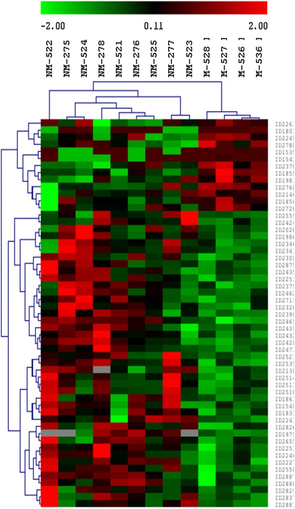
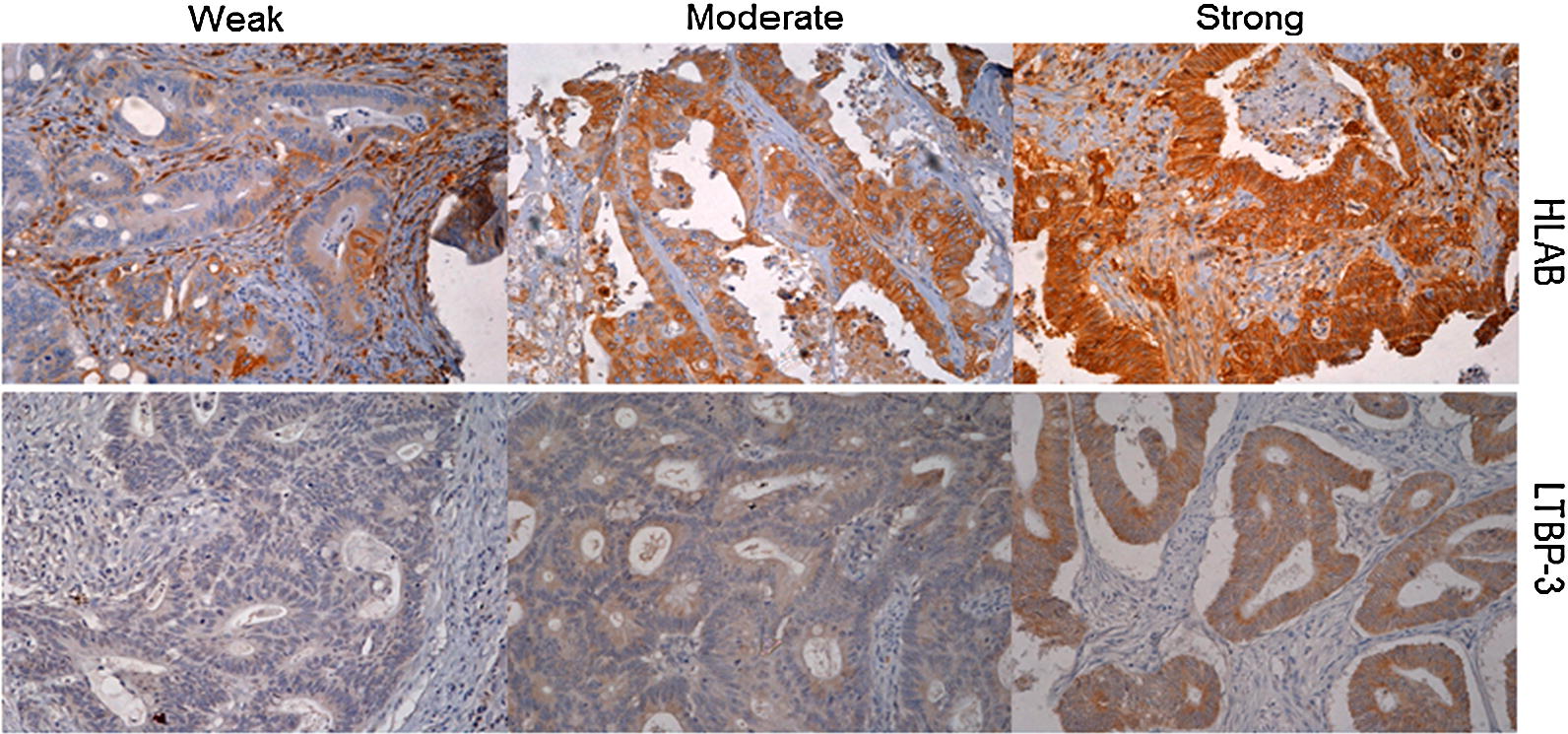
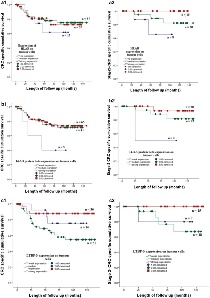
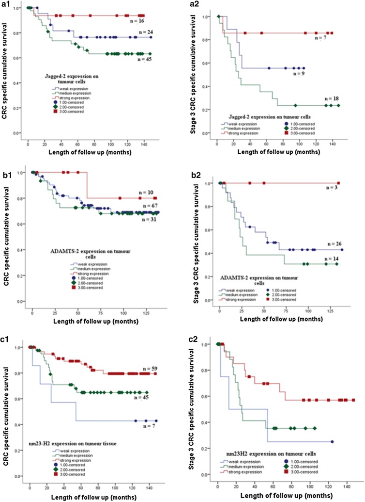
Biomarkers are urgently required to support current histological staging to provide additional accuracy in stratifying colorectal cancer (CRC) patients according to risk of spread to properly assign adjuvant chemotherapy after surgery. Chemotherapy is given to patients with stage III to reduce the risk of recurrence but is controversial in stage II patients. Up to 25% of stage II patients will relapse within 5 years after tumor removal and when this occurs cure is seldom possible. The aim of this study was to identify protein biomarkers to stratify risk of spread of CRC patients. Laser micro-dissection was used to isolate cancer cells from primary colorectal tumors of stage II patients which did or did not metastasize within 5 years after surgical resection. Protein expression differences between two groups of tumors were profiled by 2D-DIGE with saturation CyDye labeling and identified using MALDI-TOF mass spectrometry. Evaluation of protein candidates was conducted using tissue micro array (TMA) immunohistochemistry on 125 colorectal tumor tissue samples of different stages. A total of 55 differentially expressed proteins were identified. Ten protein biomarkers were chosen based on p value and ratio between non metastasized and metastazised groups and evaluated on 125 tissues using TMA immunohistochemistry. Expression of HLAB, protein 14-3-3β, LTBP3, ADAMTS2, JAG2 and NME2 on tumour cells was significantly associated with clinical parameters related to tumour progression, invasion and metastasis. Kaplan-Meier survival curve showed strong expression of six proteins was associated with good CRC specific survival. Expression of HLAB, ADAMTS2, LTBP3, JAG2 and NME2 on tumour cells, was associated with tumour progression and invasion, metastasis and CRC specific survival may serve as potential biomarkers to stratify CRC patients into low and high risk of tumour metastasis. Combined methods of laser microdissection, 2D DIGE with saturation labelling and MALDI-TOF MS proved to be resourceful techniques capable of identifying protein biomarkers to predict risk of spread of CRC to liver.

Keywords: Biomarkers; Colorectal cancer; Liver metastasis; Prognosis; Proteomics.


Figures

Fig. 1
Fig. 1
Hierarchical clustering dendrogram and heat map of differentially expressed proteins (p ≤ 0.05; fold change ≥ 1.5 or ≤ −1.5*) between the non-metastasized (NM) and metastasized (M) patient groups of colorectal cancer within 5 years after the primary tumors were removed. The dendrogram and heat map illustrate the protein expression patterns between the two groups with the segregation and alignment of proteins with similar functional groups and clustering of experimental groups with similar disease pathology. Red indicates a high relative level of protein expression, black for median value and green indicates low relative level of protein expression. *The fold change is defined as the ratio of the protein spots’ quantities of a matched protein in NM over M if its value was greater than 1, or the negative reciprocal of the ratio if its value was smaller than 1
Fig. 2
Fig. 2
Representative CRC tissues showing low, moderate and strong staining pattern of HLAB (top) and LTBP3 (bottom) proteins on tumour cells by immunohistochemistry (DAB substrate brown) (×20 objective). Sections were counterstained with hematoxylin (blue)
Fig. 3
Fig. 3
The expression of HLAB (A1) 14-3-3 protein beta (B1) and LTBP3 (C1) of the tumour cells of stage 1, 2, 3 CRC patients in a cohort of 125 tumor tissues is associated with CRC specific survival. Strong expression of HLAB, 14-3-3 protein beta and LTBP3 on cancer cells of stage 2 CRC patients showed better CRC specific survival than moderate and or weak expression of the proteins respectively (A2, B2, C2)
Fig. 4
Fig. 4
The expression of JAG2 (A1) ADAMTS2 (B1) and NME2 (C1) of the tumour cells of stage 1, 2, 3 CRC patients in a cohort of 125 tumor tissues is associated with CRC specific survival. Strong expression of JAG2, ADAMTS2 and NME2 on cancer cells of stage 3 CRC patients showed better CRC specific survival than moderate and weak expression of the proteins respectively (A2, B2, C2)

Similar articles

Cited by

References

    1. Sung JJ, Lau JY, Goh KL, Leung WK. Asia Pacific Working Group on Colorectal Cancer Increasing incidence of colorectal cancer in Asia: implications for screening. Lancet Oncol. 2005;6(11):871–876. doi: 10.1016/S1470-2045(05)70422-8. - DOI - PubMed
    1. Favoriti P, Carbone G, Greco M, Pirozzi F, Pirozzi EM, Corcione F. Worldwide burden of colorectal cancer: a review. Updates Surg. 2016;68:7–11. doi: 10.1007/s13304-016-0359-y. - DOI - PubMed
    1. Christofori G. New signals from the invasive front. Nature. 2006;441(7092):444–450. doi: 10.1038/nature04872. - DOI - PubMed
    1. Zaniboni AR, Labianca R. Adjuvant therapy for stage II colon cancer: an elephant in the living room? Ann Oncol. 2004;15(9):1310–1318. doi: 10.1093/annonc/mdh342. - DOI - PubMed
    1. Johnston PG. Stage II colorectal cancer: to treat or not to treat. Oncologist. 2005;10(5):332–334. doi: 10.1634/theoncologist.10-5-332. - DOI - PubMed