. 2019 Jan 17:156:5.
doi: 10.1186/s41065-019-0081-0. eCollection 2019.

The evolution of transcriptional repressors in the Notch signaling pathway: a computational analysis

Affiliations

The evolution of transcriptional repressors in the Notch signaling pathway: a computational analysis

Dieter Maier. Hereditas. .

Abstract

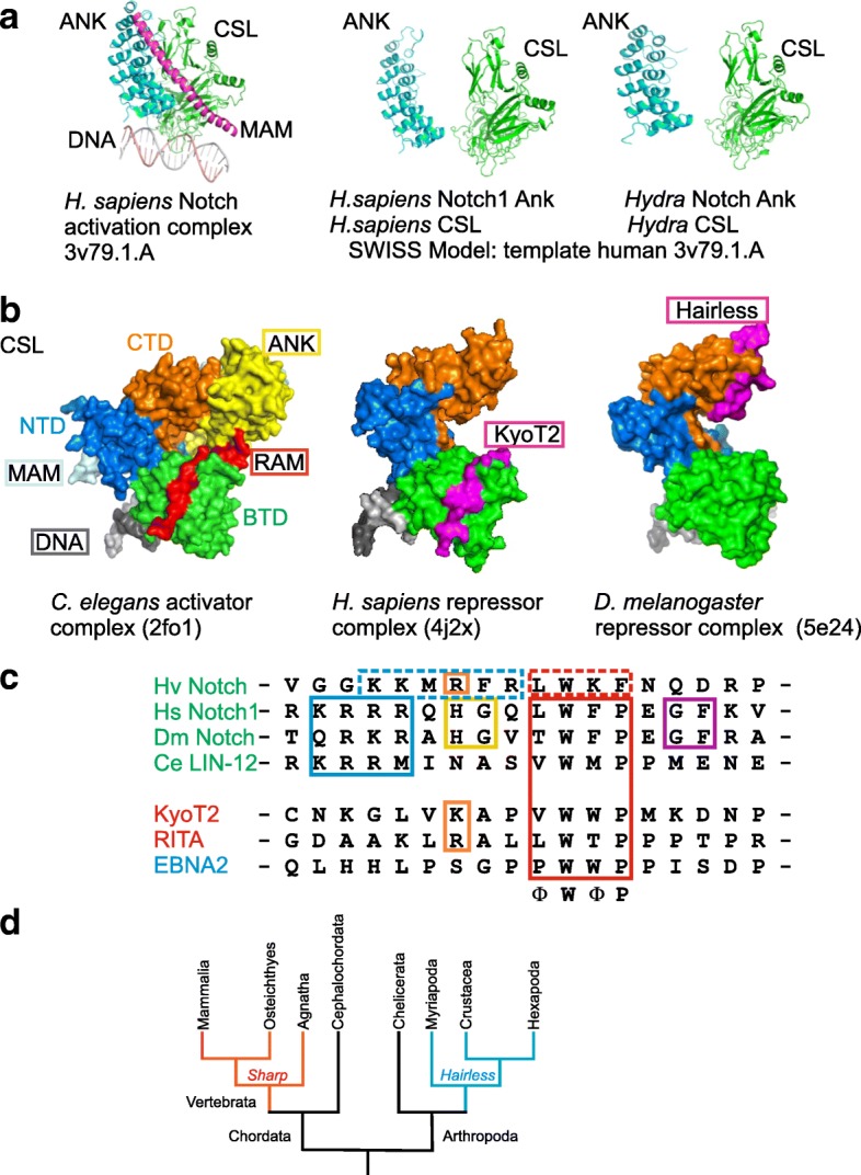
Background: The Notch signaling pathway governs the specification of different cell types in flies, nematodes and vertebrates alike. Principal components of the pathway that activate Notch target genes are highly conserved throughout the animal kingdom. Despite the impact on development and disease, repression mechanisms are less well studied. Repressors are known from arthropods and vertebrates that differ strikingly by mode of action: whereas Drosophila Hairless assembles repressor complexes with CSL transcription factors, competition between activator and repressors occurs in vertebrates (for example SHARP/MINT and KyoT2). This divergence raises questions on the evolution: Are there common ancestors throughout the animal kingdom?

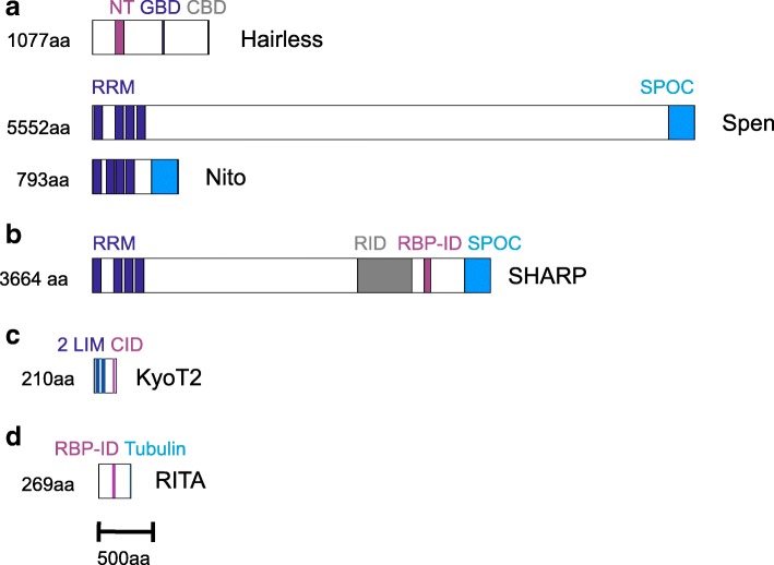
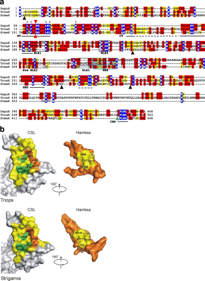
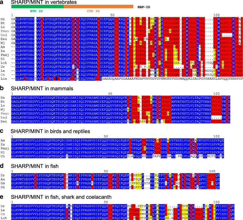
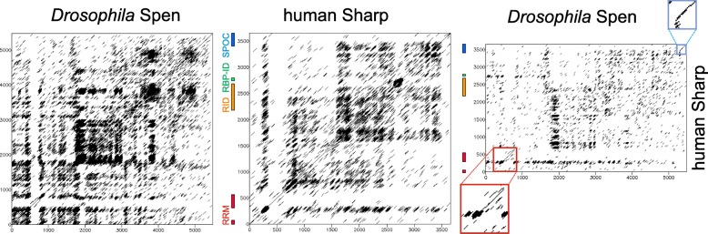
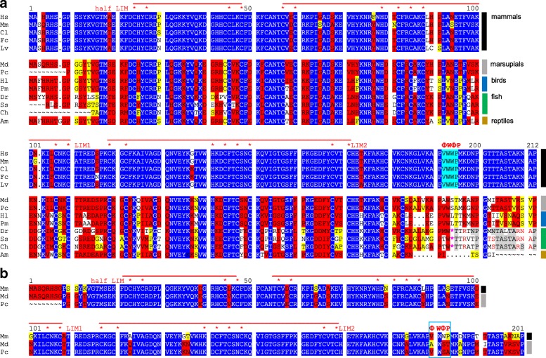
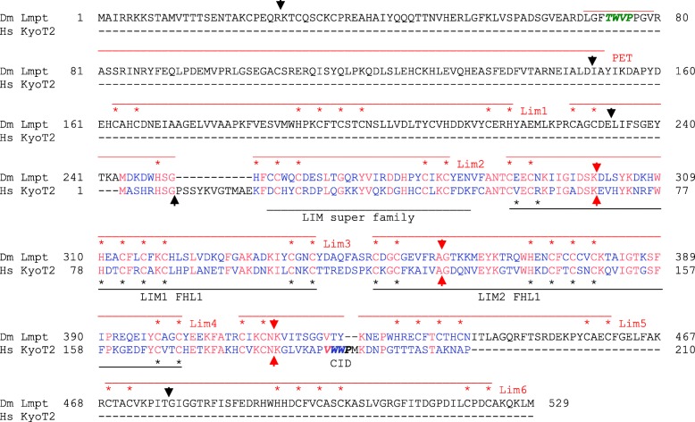
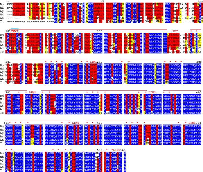
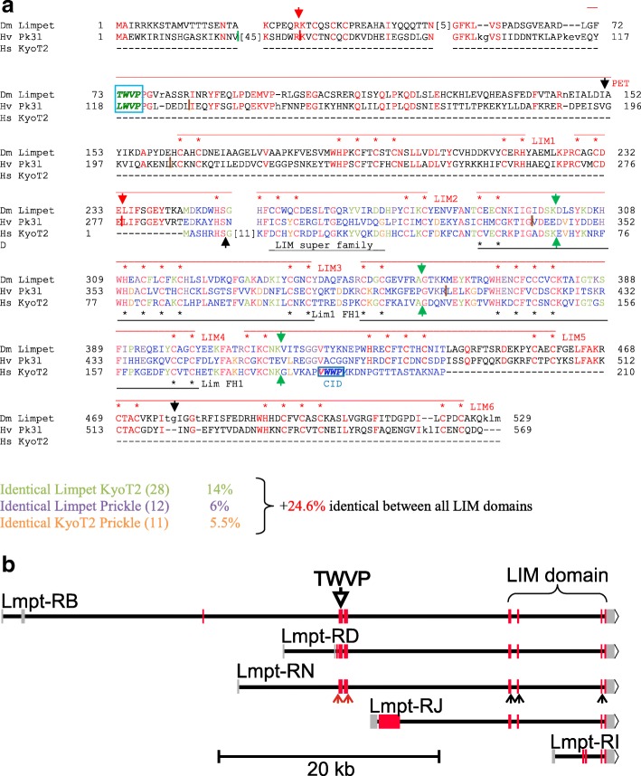
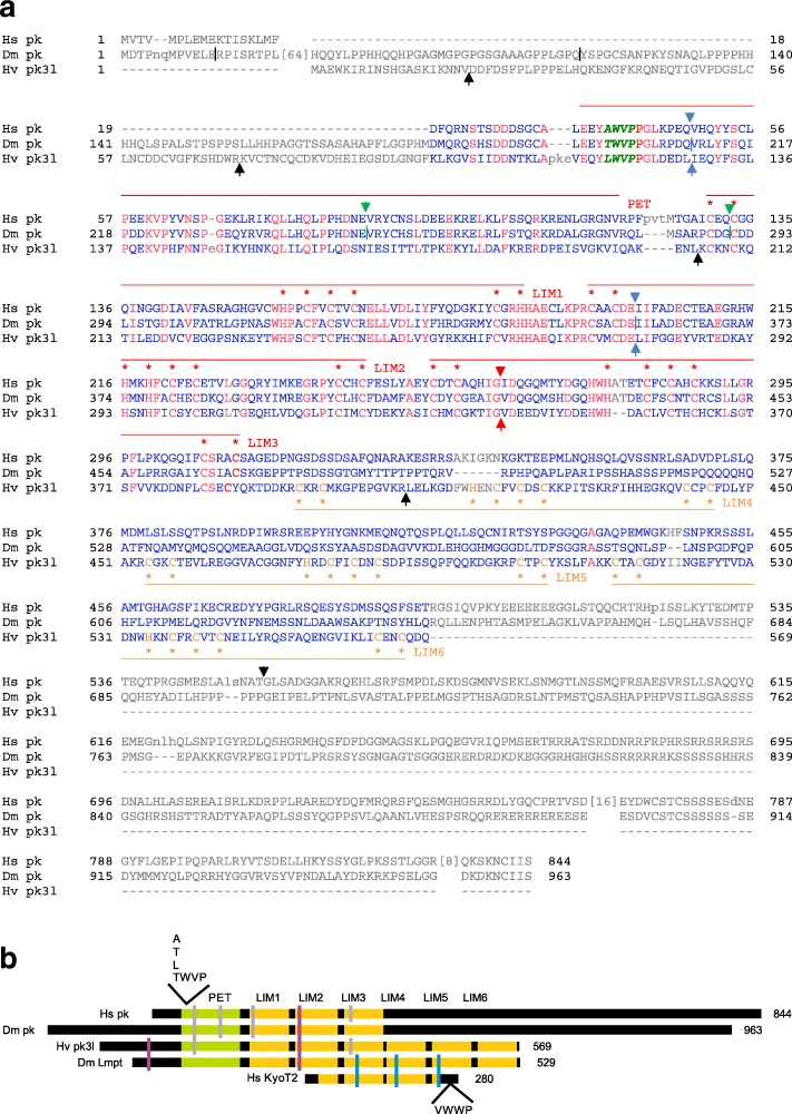
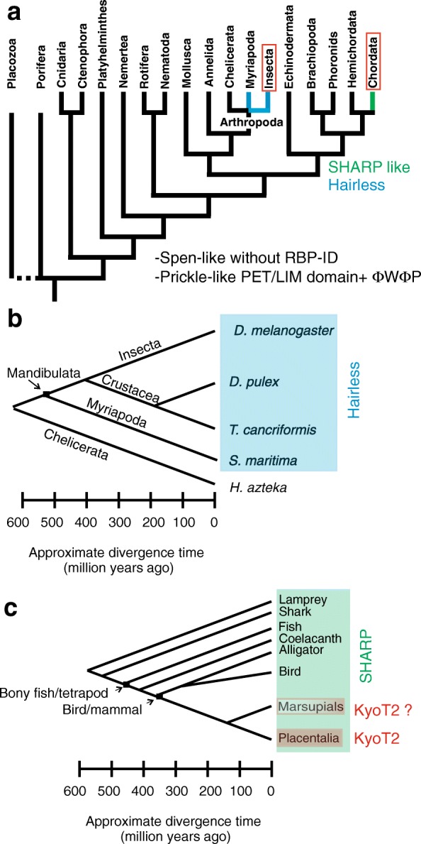
Results: Available genome databases representing all animal clades were searched for homologues of Hairless, SHARP and KyoT2. The most distant species with convincing Hairless orthologs belong to Myriapoda, indicating its emergence after the Mandibulata-Chelicarata radiation about 500 million years ago. SHARP shares motifs with SPEN and SPENITO proteins, present throughout the animal kingdom. The CSL interacting domain of SHARP, however, is specific to vertebrates separated by roughly 600 million years of evolution. KyoT2 bears a C-terminal CSL interaction domain (CID), present only in placental mammals but highly diverged already in marsupials, suggesting introduction roughly 100 million years ago. Based on the LIM-domains that characterize KyoT2, homologues can be found in Drosophila melanogaster (Limpet) and Hydra vulgaris (Prickle 3 like). These lack the CID of KyoT2, however, contain a PET and additional LIM domains. Conservation of intron/exon boundaries underscores the phylogenetic relationship between KyoT2, Limpet and Prickle. Most strikingly, Limpet and Prickle proteins carry a tetra-peptide motif resembling that of several CSL interactors. Overall, KyoT2 may have evolved from prickle and Limpet to a Notch repressor in mammals.

Conclusions: Notch repressors appear to be specific to either chordates or arthropods. Orthologues of experimentally validated repressors were not found outside the phylogenetic group they have been originally identified. However, the data provide a hypothesis on the evolution of mammalian KyoT2 from Prickle like ancestors. The finding of a potential CSL interacting domain in Prickle homologues points to a novel, very ancestral CSL interactor present in the entire animal kingdom.

Keywords: Animal kingdom; Evolution; Gene annotation; Hairless; KyoT2; Limpet; Notch repressors; Prickle; SHARP.


Conflict of interest statement

Not applicable.Not applicable.The author declares that there are no competing financial, personal, or professional interests.Springer Nature remains neutral with regard to jurisdictional claims in published maps and institutional affiliations.

Figures

Fig. 1
Fig. 1
Structure of CSL transcription complexes. a) Structure of the human Notch activator complex on DNA (PBD ID: 3v79.1A): CSL (green), Notch 1 ANK-repeats (blue) and the kinked alpha-helical domain of MAML (magenta) (left). Structure prediction of Hydra vulgaris activation complex (right), depicting Notch ANK and CSL, was done by SWISS Model using 3v79.1A as template for human Notch 1 ANK and CSL (center). b) CSL harbors three subdomains, the N-terminal domain NTD, the beta trefoil domain (BTD) and the C-terminal domain (CTD). NTD and BTD contact the DNA (grey). Left: In the activator complex (C. elegans [PDB ID: 2fo1]) Notch makes contacts to the CTD with the ANK domains (yellow), and to the BTD with its RAM domain (red). Mam (light blue) contacts BTD and ANK. Middle: The CSL-ID of KyoT2 (pink) (PDB-ID: 4J2X) makes very similar contacts as RAM with the BTD. Right: In contrast, contacts between fly Hairless (pink) and Su(H) are restricted to the CTD only (PDB ID: 5E24). c) Sequence comparison of the CSL-interacting domain from KyoT2, RITA and EBNA2 with RAM domains of Notch [Hydra vulgaris (Hv Notch), Homo sapiens (Hs Notch1), Drosophila melanogaster (Dm Notch), Caenorhabditis elegans (Ce LIN-12)]. Note lack of the typical ΦWΦP motif in Hydra Notch (Φ, any hydrophobic residue). d) Simplified phylogenetic tree of chordates and arthropods. Red: branches with SHARP coding genes, blue: with Hairless coding genes
Fig. 2
Fig. 2
Cartoons of validated Notch repressors. Notch repressor proteins characterized in Drosophila and mammals are shown to scale. Protein size is given in amino acids (aa). a) Drosophila melanogaster. Upper panel displays H protein structure with the Su(H) interacting NT box (NT), the Groucho binding domain (GBD) and the C-terminal binding protein binding domain (CBD). Split ends protein (Spen, center panel) and Spenito (Nito, lower panel) contain four RNA recognition motifs (RRM) at the N-terminus, and a Spen paralog and ortholog C-terminal (SPOC) domain. b) Homo sapiens. SHARP protein belongs to the Spen protein family; it contains in addition to the RRM and the SPOC domain also a receptor interacting domain (RID), and the RBPJ interacting domain (RBP-ID). c) Mus musculus. KyoT2 is characterized by two complete LIM domains and the CSL interacting domain (CID) d) Homo sapiens. RITA protein contains a central RBP interacting domain (RPB-ID) and a Tubulin binding motif at the C-terminus
Fig. 3
Fig. 3
Hairless orthologs from Triops and Strigamia. a Alignment of predicted Hairless orthologs from Triops cancriformis (TrcaH) and Strigamia maritima (StmaH) with the Daphnia pulex (DapuH) ortholog. Daphnia and Triops Hairless share the characterized functional domains, the Su(H) binding domain SBD which is subdivided into NT and CT box, the Groucho binding domain GBD, and the CtBP binding domain CBD. Only NT box and CBD are clearly discernable in Strigamia Hairless. The CT box and the GBD may be present with weak conservation only (dashed lines). Notably, residues known to contact the Su(H) CTD in D. melanogaster are identical in all the species (*). High confidence NLS and NES sequences are boxed, those of low confidence are dotted. Red arrowheads indicates an intron shared by all three genes. Black and blue arrowheads depict intron positions specific to Strigamia and Triops Hairless genes, respectively. Identical residues are marked in blue, highly conserved in red, conserved in yellow. b Structure predictions of the Notch repressor complexes from Triops and Strigamia, respectively, using SWISS Model and template PDB ID: 5E24 of the Drosophila complex. Left CSL CTD and right Hairless interacting domain. Potentially interacting residues are highlighted in yellow in Hairless, and green and orange in CSL
Fig. 4
Fig. 4
Comparison of SHARP/MINT proteins in vertebrates. Comparison of SHARP/MINT orthologues in vertebrates; shown is the alignment of the RBP-ID and C-terminally adjacent amino acids. Identical residues are marked in blue identical, highly conserved in red, conserved in yellow. a Alignment over the whole vertebrate tree, b) alignment of mammals, c) alignment of birds and reptiles, d) alignment of fish, e) alignment of zebra fish with chondrichthyes (cartilaginous fish) and coelacanth. Human RBP-ID, shown to interact with CSL BTD and CSL CTD, is highly conserved in all vertebrates except the arctic lamprey, where it ends a bit short. Also C-terminally adjacent sequences except of lamprey (italic) are conserved, which becomes apparent in the comparison of the group specific alignments. The rattlesnake, however, is distinctly different from alligator and birds in this region. Mammals: Hs: Homo sapiens (man), Bt: Bos taurus (cattle), Lv: Lipotes vexillifer (chinese river dolphin), Mj: Manis javanica (malayan pangolin), Pman: Peromyscus maniculatus (great tit), Eed: Elephantulus edwardii (cape elephant shrew), Eeu: Erinaceus europaeus (european hedgehog), Reptile and birds: Am: Alligator mississippiensis (alligator), Za: Zonotrichia albicollis (sparrow), Pmaj: Parus major (great tit), Hl: Haliaeetus leucocephalus (bald eagle), Ch: Crotalus horridus (timber rattlesnake), Fish: Dr.: Danio rerio (zebrafish), Aa: Anguilla Anguilla (eel), Gm: Gadus morhua (ghostshark), Ok: Oncorhynchus kisutch (coho salmon), Le: Leucoraja erinacea (little skate), Cm: Callorhinchus milii (ghostshark), Lca: Lethenteron camtschaticum (lamprey), Lch: Latimeria chalumnae (coelacanth) See also Additional file 3 Dataset 2 for common names
Fig. 5
Fig. 5
Dotplot analysis of Spen and SHARP. The dotplot analysis of Drosophila Spen and human SHARP shows typical repetition patterns that reflect the accumulation of single amino acids. A dotplot comparison Drosophila Spen with human SHARP reveals clear similarities only in the RRM and SPOC domains shown enlarged (red box, RRM and blue box, SPOC domain). The RID (orange) and RBP-ID (green) domains in SHARP are indicated as well
Fig. 6
Fig. 6
Alignment of KyoT2 homologues from vertebrates. a Only placental mammals contain a KyoT2 protein with conserved CSL binding motif (ΦWΦP, cyan). Already in marsupial (koala P. cinereus and opossum M. domestica) or birds (eagle H. leudocephalus and tit P. major) the motif is changed, but an open reading frame still remains. In fish, a stop codon (*) occurs exactly within the motif, however, sequences C-terminally translate into many conserved residues (grey) compared to mammals. All proteins consist of two and a half LIM domains (red lines), each containing two tandemly repeated zinc fingers. Asterisks indicate zinc binding residues. Blue are identical, red are highly conserved and yellow are related residues. Note changes in marsupials. b Alignment of mouse KyoT2 with marsupial koala and opossum. Note high overall conservation. The ΦWΦP tetra-peptide motif is highlighted Placental mammals (black box): Hs: Homo sapiens (man), Mm: Mus musculus (mouse), Cl: Canis lupus (wolf), Fc: Felis catus (cat), Lv: Lipotes vexillifer (chinese river dolphin); Marsupials (grey box): Pc: Phascolarctos cinereus (koala), Md: Monodelphis domestica (opossum). Birds (blue box): Hl: Haliaeetus leucocephalus (bald eagle), Pm: Parus major (great tit). Fish (green box): Dr.: Danio rerio (zebrafish), Ss: Salmon solar (atlantic salmon), Ch: Clupea harengus (herring). Reptiles (brown box): Am: Alligator mississippiensis.
Fig. 7
Fig. 7
Drosophila Limpet is a homolog of human KyoT2. D. melanogaster Limpet protein harbors a PET domain, followed by six LIM domains. The alignment with human KyoT2 (also named Fhl1C) reveals surprising similarity between the LIM domains of KyoT2 (black labelling) and LIM domains two to four of Drosophila Limpet (isoform PN, red labelling). Intriguingly, three identical intron positions are present within this region (red arrows). Black arrows mark intron positions specific to Limpet. The CSL binding domain of KyoT2 (CID, italics) is not present in Limpet at a corresponding position. A similar tetra-peptide motif TWVP (italic green), however, is located at the N-terminus of the PET domain. Red residues are identical between the two proteins, blue are not conserved, black absent in one of the two species
Fig. 8
Fig. 8
Limpet orthologs in insects. Limpet orthologs are only found in higher insects. Highest conservation is seen in the LIM domains with many identical residues (blue), whereas the PET domain and the N-terminus are less well conserved. The tetra-peptide Lmpt-motif TWVP (italic, boxed) is present in all aligned sequences at identical position, with conservative variations seen in Tribolium castaneum (Tc) and Apis dorsata (Ad). Flies, Dm: Drosophila melanogaster, Md: Musca domestica, Ag: Anopheles gambiae, Aa: Aedes aegypti, Bee, Ad: Apis dorsata. Beetle, Tc: Tribolium castaneum (see also Additional dataset 2 for common names)
Fig. 9
Fig. 9
Alignment of fly Limpet, human KyoT2 and Hydra Prickle 3 like. a The alignment shows good conservation in the LIM domain between all three proteins. Best identity score in the LIM domain is between KyoT2 and Limpet (38.6%), followed by Limpet and Prickle 3 like (30.6%) and KyoT2 and Prickle 3 like (30.1%). Limpet and Prickle 3 like share considerable conservation over the entire length; the TWVP tetra-peptide motif is present at the identical position in the PET domain (italic green, boxed). Moreover, the two share conserved intron positions (red arrows and dashes). Green arrows indicate identical intron positions between Limpet and KyoT2. Black arrows and dashes mark intron positions specific to Hydra Prickle 3 like. Red residues are identical in all three proteins, green identical between Limpet and KyoT2, pink identical between Limpet and Prickle 3 like, and orange identical between KyoT2 and Prickle 3 like, blue are not conserved. b Scheme of the genomic organization of the D. melanogaster Limpet gene with representative transcripts (adapted from flybase). 15 transcripts are predicted, however, with 8 different open reading frames that fall into three classes: One encoding PET and LIM domain proteins (RB, RD and RN are shown as examples), one encoding LIM domain only proteins (RI, and RJ are shown as example), and the third containing neither (not shown). The exon encoding the PET domain with the TWVP tetra-peptide is indicated by the open arrow. The small downstream exons encode the LIM domain. Red arrows indicate exon/intron boundaries conserved between Limpet and Prickle 3 like, black arrows indicate exon/intron boundaries conserved between Limpet and KyoT2
Fig. 10
Fig. 10
Conservation of Prickle like proteins. a Alignment of human Prickle homolog 2 (HS pk; GenBank AAI19003.1), Drosophila melanogaster Prickle isoform A (Dm pk; flybase) and Hydra vulgaris Prickle 3 like (Hv pk3l). All three share the PET domain and the first three LIM domains. In this part many residues are identical (red), blue residues are not conserved. The Hydra protein has three additional LIM domains, whereas the human and fly Prickle proteins extend C-terminally with no further similarities or discerned motifs. Reduced stringency conditions were used to allow alignment of LIM domain three. Notably, a tetra-peptide motif TMVP with high similarity to the Lmpt motif is shared by all Prickle proteins at the corresponding position (italic green) in the PET domain. Blue arrows indicate intron positions identical in all genes, green identical in human and Drosophila and red identical between human and Hydra. Black arrows and dashes present species specific introns. b Schematic comparison of Prickle (pk), Prickle 3 like (pk3l) and Limpet (Lmpt) proteins from human (Hs), Drosophila (Dm) and Hydra (Hv). PET and LIM domains are color coded green and yellow, respectively. Intron positions are indicated by dashes: purple conserved in Limpet and prickle, blue conserved between KyoT2 and Drosophila Limpet and grey conserved in prickle. Only conserved intron positions are shown. Positions of defined CID motif in KyoT2 (VWWP) and potential interacting motifs (TWVP like) in the others are indicated
Fig. 11
Fig. 11
Phylogeny of CSL transcription complexes. a Phylogenetic tree of the various animal clades from this study. Notch repressors Hairless, SHARP, and KyoT2 were not found outside the clade, where they have been originally identified. The Spen-like and Prickle-like family of proteins, however, is present in all the studied metazoan animals. b Phylogeny of Hairless evolution. Hairless can be traced back to Myriapoda that diverged from dipteran flies approximately 500 million years ago, i.e. after the Mandibulata-Chelicerata radiation. c Phylogeny of SHARP/MINT and KyoT2 evolution. SPEN-like proteins containing a RBPJ interacting domain (i.e. SHARP) are only found in vertebrates starting with arctic lampreys as example for Cyclostomata. Lampreys are considered living fossils that may not have changed morphologically the past 500 million years, but diverged about 600 million years back from mammals. The KyoT2 co-repressor, however, is only found in placental mammals. The CSL interacting motif in marsupial KyoT2 is quite diverged, presumably impeding the binding to CSL

Similar articles

Cited by

References

    1. Artavanis-Tsakonas S, Rand MD, Lake RJ. Notch signaling: cell fate control and signal integration in development. Science. 1999;284(5415):770–776. doi: 10.1126/science.284.5415.770. - DOI - PubMed
    1. Kopan R, Ilagan MXG. The canonical notch signaling pathway: unfolding the activation mechanism. Cell. 2009;137:216–233. doi: 10.1016/j.cell.2009.03.045. - DOI - PMC - PubMed
    1. Schlatter R, Maier D. The enhancer of split and Achaete-Scute complexes in Drosophilids are derived from simple ur-complexes preserved in mosquito and honeybee. BMC Evol Biol. 2005;5:67. doi: 10.1186/1471-2148-5-67. - DOI - PMC - PubMed
    1. Käsbauer T, Towb P, Alexandrova O, David CN, Dall'Armi E, Staudigl A, Stiening B, Böttger A. The notch signaling pathway in the cnidarian Hydra. Dev Biol. 2007;303:376–390. doi: 10.1016/j.ydbio.2006.11.022. - DOI - PubMed
    1. Dexter JS. The analysis of a case of continuous variation in Drosophila by a study of its linkage relations. Am Nat. 1914;48:712–758. doi: 10.1086/279446. - DOI

Substances

LinkOut - more resources