

Interaction between a 3D collagen matrix used for periodontal soft tissue regeneration and T-lymphocytes: An in vitro pilot study
- PMID: 30679964
- PMCID: PMC6327677
- DOI: 10.3892/etm.2018.6979
Interaction between a 3D collagen matrix used for periodontal soft tissue regeneration and T-lymphocytes: An in vitro pilot study
Abstract
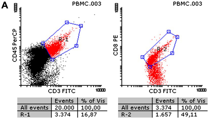
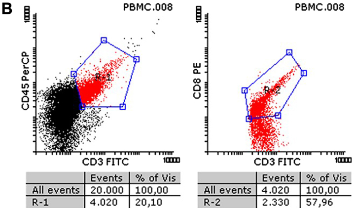
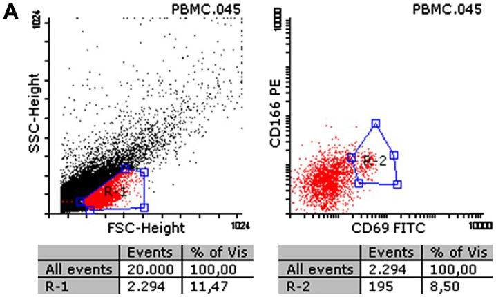
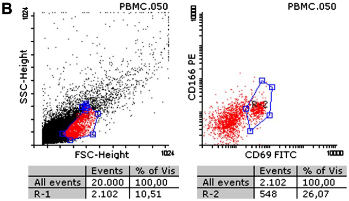
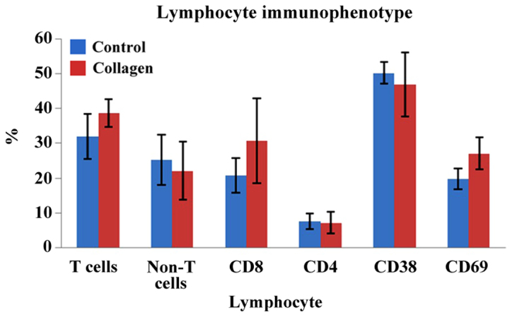
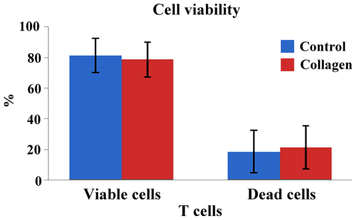
Previous experimental models showed that activation of the immune system, particularly T cells, is required for optimal healing following wounds or surgery in the oral cavity. Therefore, studies to explore the interactions between the immune system and the collagen matrix are mandated. The specific aim of the present study was to analyze the interactions between T lymphocytes and a resorbable three-dimensional (3D) collagen matrix routinely used for soft tissue regeneration during periodontal surgery. Peripheral venous blood samples were collected from five patients. Following Ficoll-Paque separation, mononuclear cells were grown on fully resorbable 3D collagen matrices for 5 days. Lymphocytes were analyzed by flow cytometry for different surface markers, including CD4, CD8, CD38 and CD69. Cell viability and late apoptosis/necrosis were assessed in each group using an apoptosis assay based on Annexin V/propidium iodide staining. After 5 days in contact with the collagen matrix, the T cells expressed different surface markers. The overall T cell population increased significantly in the collagen matrix group compared to the respective controls (31.9±6.5 vs. 38.7±3.8%). CD8 and CD69 also increased significantly compared to their controls (CD69: 19.7±3.0 vs. 27.1±4.5% for collagen vs. control groups). At the same time, CD4 and CD38 expression was similar in both groups. Viability and apoptosis/necrosis were also identical in the samples and controls. These results show that the interaction between the collagen matrix and the immune cells stimulated activation of T cells and did not impair the healing process.
Keywords: T-lymphocytes; apoptosis; collagen matrix; oral mucosa; wound healing.
Figures
References
-
- Nevins M, Nevins ML, Kim SW, Schupbach P, Kim DM. The use of mucograft collagen matrix to augment the zone of keratinized tissue around teeth: A pilot study. Int J Periodontics Restorative Dent. 2011;31:367–373. - PubMed
-
- Hall WB, Lundergan WP. Free gingival grafts. Current indications and techniques. Dent Clin North Am. 1993;37:227–242. - PubMed
-
- Tonetti MS, Jepsen S. Working Group 2 of the European Workshop on Periodontology: Clinical efficacy of periodontal plastic surgery procedures: Consensus report of Group 2 of the 10th European Workshop on Periodontology. J Clin Periodontol. 2014;41(Suppl 15):S36–S43. doi: 10.1111/jcpe.12219. - DOI - PubMed
LinkOut - more resources
Full Text Sources
Research Materials