

In vivo confocal laser scanning microscopy imaging of skin inflammation: Clinical applications and research directions
- PMID: 30679966
- PMCID: PMC6327452
- DOI: 10.3892/etm.2018.6981
In vivo confocal laser scanning microscopy imaging of skin inflammation: Clinical applications and research directions
Abstract
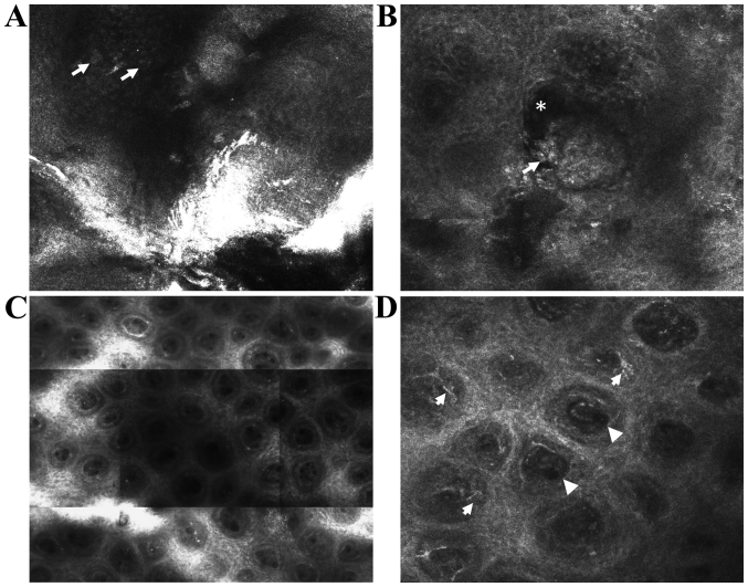
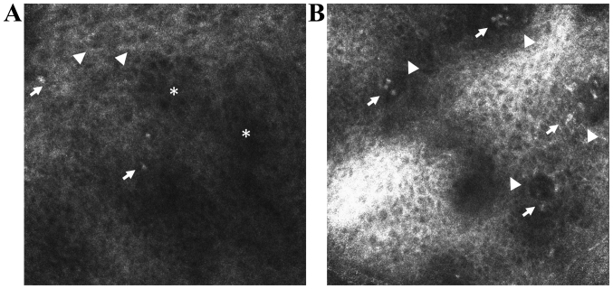
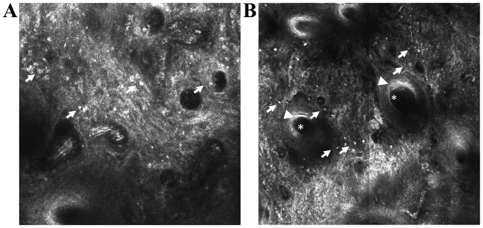
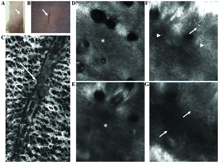
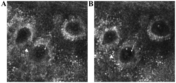
In vivo confocal laser scanning microscopy (CLSM) is a novel imaging technique that provides noninvasive, morphological characterization of skin structures with a resolution that is very close to that of light microscopy. Moreover, as it allows repeated imaging of the same skin area at different time-points, it is an excellent method for monitoring disease course, response to treatment or specific stimuli and a path to study dynamic phenomena in real-time. To date, two different variants of in vivo CLSM have been authorized in dermatological field, namely the reflectance confocal microscopy predominantly for clinical diagnosis and the fluorescence confocal microscopy mainly for research purposes. This study describes the principles of in vivo CLSM technique, its role in the diagnosis and monitoring of inflammatory skin diseases, as well as some promising research directions to study the dynamics of skin inflammation using this method. In vivo CLSM evaluation of inflammatory dermatoses and of the skin inflammatory component in various diseases has an undoubted potential with broad applications ranging from clinical, morphological to experimental, functional studies involving the skin.
Keywords: in real-time; in vivo; reflectance confocal microscopy; skin inflammation.
Figures
References
-
- Ghiţă MA, Căruntu C, Rosca AE, Căruntu A, Moraru L, Constantin C, Neagu M, Boda D. Real-time investigation of skin blood flow changes induced by topical capsaicin. Acta Dermatovenerol Croat. 2017;25:223–227. - PubMed
Publication types
LinkOut - more resources
Full Text Sources