

Role of modern imaging techniques for the in vivo diagnosis of lichen planus
- PMID: 30679973
- PMCID: PMC6327670
- DOI: 10.3892/etm.2018.6974
Role of modern imaging techniques for the in vivo diagnosis of lichen planus
Abstract
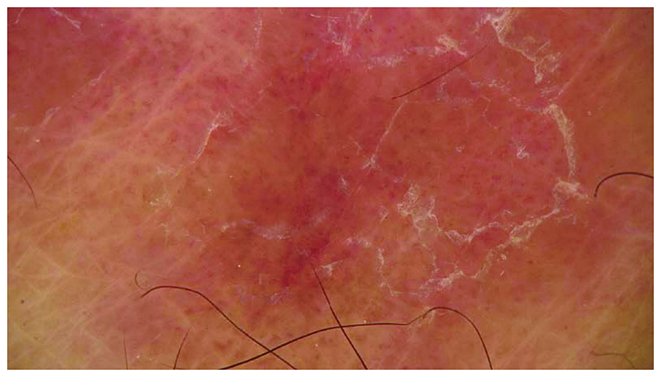
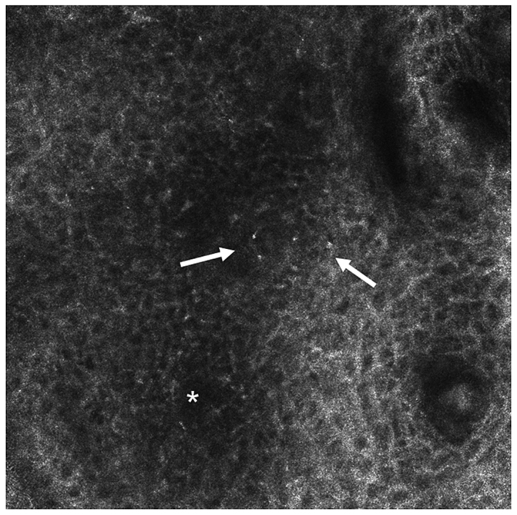
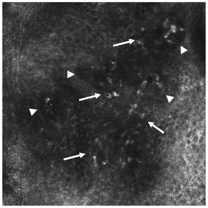
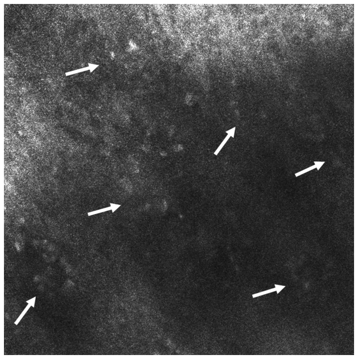
Lichen planus (LP) is a chronic inflammatory skin disease that can sometimes affect mucosal surfaces, with unknown pathogenesis, even though it appears to be an autoimmune disease. The diagnosis of lichen planus is usually based on histopathological examination of the lesions. Nowadays, the classical invasive diagnostic methods are replaced by modern non-invasive techniques. In this review, we present the main non-invasive imaging methods (dermoscopy, reflectance confocal microscopy, optical coherence tomography, ultrasound and diffuse reflection spectrophotometry) used in the diagnosis and therapeutic monitoring of lichen planus. Dermoscopy is a non-invasive method initially used for diagnosis of pigmented tumors but now is used also for inflammatory and infectious skin diseases. In lichen planus, the dermoscopy increases the accuracy of diagnosis, avoids skin biopsies commonly used and can be useful in the therapeutic monitoring by repeated investigation at different stages of treatment. Reflectance confocal microscopy (RCM) is a novel non-invasive imaging technique that is prevalently used for the diagnosis of skin tumors and inflammatory skin diseases. This technology has been mostly employed for bedside, real-time microscopic evaluation of psoriasis, lichen planus, contact dermatitis, revealing specific confocal features to support clinical diagnosis and assist with patient management. Optical coherence tomography (OCT) is an emergent imaging technique, developed over the last decade, based on the interaction of the infrared radiation (900-1,500 nm) and the living tissues. A limited information exists on the benefits of OCT technology for the in vivo diagnosis of LP but could be a useful auxiliary tool in the in vivo differential diagnosis, especially in clinical equivocal settings like mucosal lesions, and in monitoring the response to treatment. Our review shows the possibility of using modern imaging techniques for the in vivo diagnosis and also for evaluation of the treatment response.
Keywords: dermoscopy; diffuse reflection spectrophotometry; lichen planus; opticalcoherence tomography; reflectance confocal microscopy; ultrasound.
Figures
Similar articles
-
Role of In Vivo Reflectance Confocal Microscopy in the Analysis of Melanocytic Lesions.Acta Dermatovenerol Croat. 2018 Apr;26(1):64-67. Acta Dermatovenerol Croat. 2018. PMID: 29782304 Review.
-
Comparison of dermoscopy and reflectance confocal microscopy accuracy for the diagnosis of psoriasis and lichen planus.Skin Res Technol. 2022 May;28(3):480-486. doi: 10.1111/srt.13158. Epub 2022 Mar 28. Skin Res Technol. 2022. PMID: 35347763 Free PMC article.
-
Clinical application of optical coherence tomography for the imaging of non-melanocytic cutaneous tumors: a pilot multi-modal study.J Med Life. 2010 Oct-Dec;3(4):381-9. J Med Life. 2010. PMID: 21254735 Free PMC article. Clinical Trial.
-
Pilot study on reflectance confocal microscopy imaging of lichen planus: a real-time, non-invasive aid for clinical diagnosis.J Eur Acad Dermatol Venereol. 2012 Oct;26(10):1258-65. doi: 10.1111/j.1468-3083.2011.04279.x. Epub 2011 Sep 29. J Eur Acad Dermatol Venereol. 2012. PMID: 21958425
-
Reflectance Confocal Microscopy for Inflammatory Skin Diseases.Actas Dermosifiliogr. 2016 Oct;107(8):631-9. doi: 10.1016/j.ad.2016.01.010. Epub 2016 Mar 17. Actas Dermosifiliogr. 2016. PMID: 26996333 Review. English, Spanish.
Cited by
-
In vivo Diagnosis of Primary Cutaneous Amyloidosis -the Role of Reflectance Confocal Microscopy.Diagnostics (Basel). 2019 Jun 27;9(3):66. doi: 10.3390/diagnostics9030066. Diagnostics (Basel). 2019. PMID: 31252549 Free PMC article.
-
Looking into the Skin in Health and Disease: From Microscopy Imaging Techniques to Molecular Analysis.Int J Mol Sci. 2023 Sep 6;24(18):13737. doi: 10.3390/ijms241813737. Int J Mol Sci. 2023. PMID: 37762038 Free PMC article.
-
Imaging techniques in the diagnosis and monitoring of psoriasis.Exp Ther Med. 2019 Dec;18(6):4974-4980. doi: 10.3892/etm.2019.7957. Epub 2019 Aug 29. Exp Ther Med. 2019. PMID: 31819765 Free PMC article. Review.
-
High serum level of interleukin-6 is linked with dyslipidemia in oral lichen planus.Exp Ther Med. 2021 Sep;22(3):987. doi: 10.3892/etm.2021.10419. Epub 2021 Jul 13. Exp Ther Med. 2021. PMID: 34345269 Free PMC article.
-
A Retrospective Study of the Diagnostic Accuracy of In Vivo Reflectance Confocal Microscopy for Basal Cell Carcinoma Diagnosis and Subtyping.J Clin Med. 2019 Apr 3;8(4):449. doi: 10.3390/jcm8040449. J Clin Med. 2019. PMID: 30987174 Free PMC article.
References
Publication types
LinkOut - more resources
Full Text Sources