Review
. 2019 Jan 17:6:1.
doi: 10.1186/s40673-018-0095-9. eCollection 2019.

Slow saccades in cerebellar disease

Affiliations
Review

Slow saccades in cerebellar disease

Kelsey Jensen et al. Cerebellum Ataxias. .

Abstract

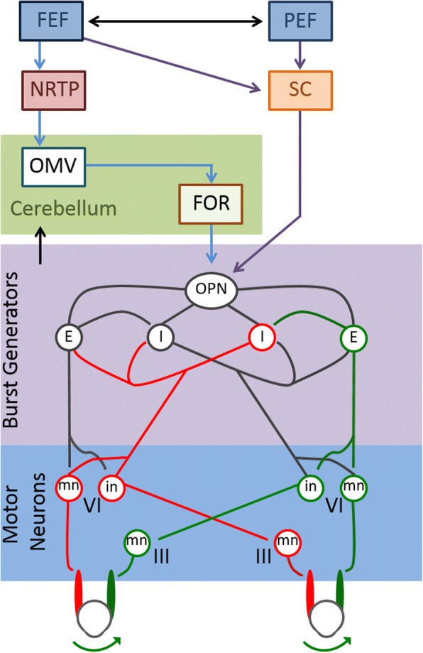
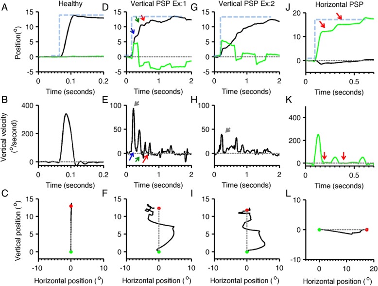
Eye movements are frequently considered diagnostic markers indicating involvement of the cerebellum. Impaired amplitude of saccades (saccade dysmetria), impaired gaze holding function (horizontal or downbeat nystagmus), and interrupted (choppy) pursuit are typically considered hallmarks of cerebellar disorders. While saccade dysmetria is a frequently considered abnormality, the velocity of saccades are rarely considered part of the constellation of cerebellar involvement. Reduced saccade velocity, frequently called "slow saccades" are typically seen in a classic disorder of the midbrain called progressive supranuclear palsy. It is also traditionally diagnostic of spinocerebellar ataxia type 2. In addition to its common causes, the slowness of vertical saccades is not rare in cerebellar disorders. Frequently this phenomenology is seen in multisystem involvement that substantially involves the cerebellum. In this review we will first discuss the physiological basis and the biological need for high saccade velocities. In subsequent sections we will discuss disorders of cerebellum that are known to cause slowing of saccades. We will then discuss possible pathology and novel therapeutic strategies.

Keywords: Burst neurons; Degenerative disorder; Dysmetria; Reciprocal innervation.


Conflict of interest statement

Kelsey Jensen (kmj53@case.edu), Sinem Balta Beylergil (sxb818@case.edu), Aasef G. Shaikh (aasefshaikh@gmail.com). Neurological Institute, University Hospitals, Cleveland, OH. Department of Neurology, Case Western Reserve University, Cleveland, OH. Neurology Service, Louis Stokes Cleveland VA Medical Center, Cleveland, OH.Not applicable - this is a review paper.This is a review paper, hence not applicable.The authors declare that they have no competing interests.Springer Nature remains neutral with regard to jurisdictional claims in published maps and institutional affiliations.

Figures

Fig. 1
Fig. 1
Neural circuitry involved in the generation of visually guided saccades. The frontal eye field (FEF) and parietal eye field (PEF) both send projections to the superior colliculus (SC), which in turn directly communicates with omniopause neurons (OPN) in the midline pons. The FEF also projects to the nucleus reticularis tegmenti pontis (NRTP) in the midbrain. This projects to the oculomotor vermis in the cerebellar cortex, which sends inhibitory fibers to the deep cerebellar fastigial oculomotor (FOR) nucleus. The FOR also communicates with OPN. The OPN is responsible for tonic inhibition of excitatory (E) and inhibitory (I) burst neurons, which results in steady fixation of gaze. When the OPN is inhibited during saccade generation, the excitatory and inhibitory burst neurons fire more rapidly. Excitatory burst neurons synapse on the abducens motoneurons (mn) and internuclear neurons (in) in the ipsilateral abducens nucleus. The internuclear neurons of the side receiving excitatory input project to the contralateral abducens motoneuronsmedial rectus subgroup of the oculomotor nucleus. Ipsilateral inhibitory burst neurons project to the contralateral abducens which inhibits movement of the opposing muscles. This results in the rapid, coordinated movement of gaze to a target object
Fig. 2
Fig. 2
The visually guided saccades from healthy subject and PSP patient are compared. Panel a illustrates normal visually guided vertical saccade from a healthy subject. Panels d,g depict two examples of visually guided vertical saccades from the same PSP subject. Panel j depicts eye positions during horizontal saccade. In panels (a,d,g,j) the eye position is plotted on the y-axis while x-axis depicts corresponding time in seconds. Black line represents the vertical eye position, while green traces depict horizontal eye position. Grey dashed line is the baseline, i.e. the straight-ahead position, while blue dashed line depicts the desired position. The arrows depict interruption in the saccades, in one type of interruption (blue arrow) the eyes continue to move at slower velocity during interruption, while in other type (green arrow) the slower eye movement in the opposite direction. The third type of interruption (red arrow) is where the eye movements completely stop during the interruption. Panels b,e, h,k depict eye velocity. Panel B depicts eye velocity of normal visually guided saccade recorded from the healthy subject, while panels (e,h) depict vertical eye velocity during vertical saccade in PSP. Green line in panel K illustrates normal horizontal eye velocity during horizontal saccade in PSP. In these subplots the eye velocity is plotted on y-axis while x-axis illustrates corresponding time. Red arrow illustrates interruption in saccade when eye velocity was zero, green arrow is when eye moved at slower velocity in the opposite direction, blue arrow is when eyes moved in the same direction at slower velocity. Panels c,f,i,l depict trajectories of horizontal and vertical saccades. Panel C shows the normal saccade from the healthy subject, while panel (f,i) are vertical saccades in PSP, and panel l is the horizontal saccade in PSP. In these plots the green dot depicts the start point, while the red dot is the stop point; grey dashed line is the desired path of the eye movement. Vertical saccades have curved and serpentine path depicting the irregularity in the trajectory. Such curvature is also present in the horizontal saccade but to much lesser extent (adapted from Shaikh et al. [121])

References

    1. Munoz DP. Progress in Brain Research. 2002. Commentary: saccadic eye movements: overview of neural circuitry; pp. 89–96. - PubMed
    1. Pierrot-Deseilligny C, Milea D, Müri RM. Eye movement control by the cerebral cortex. Curr Opin Neurol. 2004;17(1):17–25. doi: 10.1097/00019052-200402000-00005. - DOI - PubMed
    1. Fox PT, Fox JM, Raichle ME, Burde RM. The role of cerebral cortex in the generation of voluntary saccades: a positron emission tomographic study. J Neurophysiol. 1985;54(2):348–369. doi: 10.1152/jn.1985.54.2.348. - DOI - PubMed
    1. Pierrot-deseilligny CH, Rivaud S, Gaymard B, Agid Y. Cortical control of reflexive visually-guided saccades. Brain. 1991;114(3):1473–1485. doi: 10.1093/brain/114.3.1473. - DOI - PubMed
    1. Berman RA, Colby CL, Genovese CR, Voyvodic JT, Luna B, Thulborn KR, et al. Cortical networks subserving pursuit and saccadic eye movements in humans: an FMRI study. Hum Brain Mapp. 1999;8(4):209–225. doi: 10.1002/(SICI)1097-0193(1999)8:4<209::AID-HBM5>3.0.CO;2-0. - DOI - PMC - PubMed