. 2019 Mar;7(3):e556.
doi: 10.1002/mgg3.556. Epub 2019 Jan 24.

Interaction of germline variants in a family with a history of early-onset clear cell renal cell carcinoma

Affiliations

Interaction of germline variants in a family with a history of early-onset clear cell renal cell carcinoma

Emmanuelle Nicolas et al. Mol Genet Genomic Med. 2019 Mar.

Abstract

Background: Identification of genetic factors causing predisposition to renal cell carcinoma has helped improve screening, early detection, and patient survival.

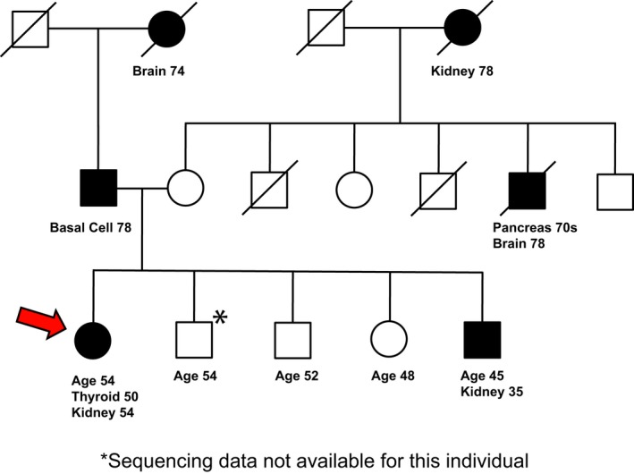
Methods: We report the characterization of a proband with renal and thyroid cancers and a family history of renal and other cancers by whole-exome sequencing (WES), coupled with WES analysis of germline DNA from additional affected and unaffected family members.

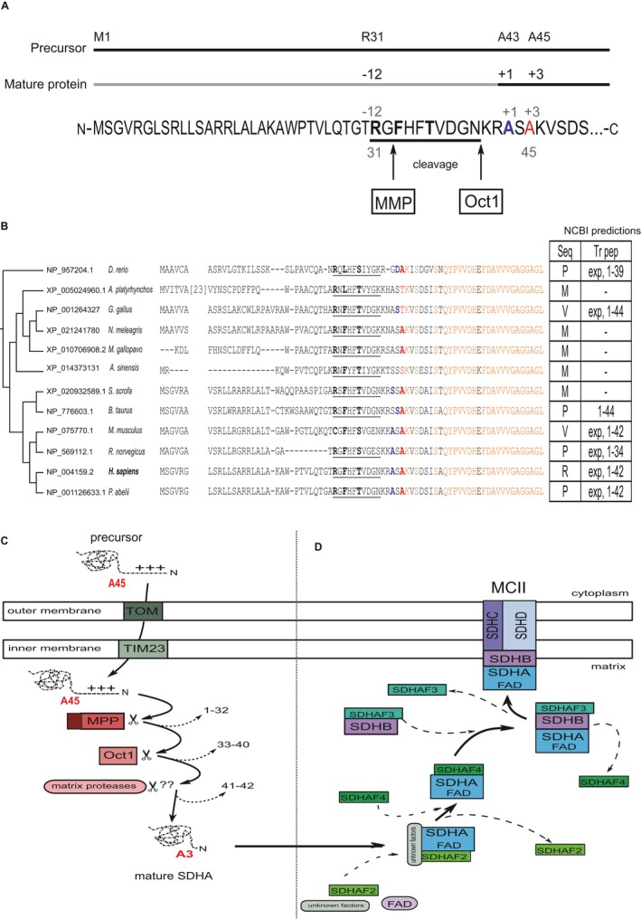
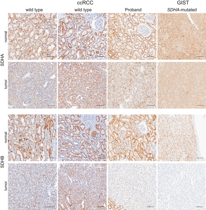
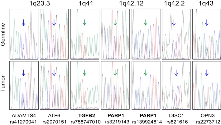
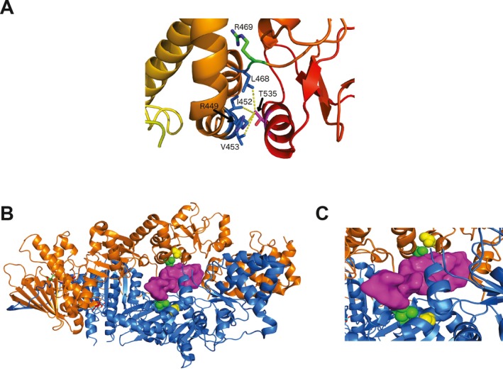
Results: This work identified multiple predicted protein-damaging variants relevant to the pattern of inherited cancer risk. Among these, the proband and an affected brother each had a heterozygous Ala45Thr variant in SDHA, a component of the succinate dehydrogenase (SDH) complex. SDH defects are associated with mitochondrial disorders and risk for various cancers; immunochemical analysis indicated loss of SDHB protein expression in the patient's tumor, compatible with SDH deficiency. Integrated analysis of public databases and structural predictions indicated that the two affected individuals also had additional variants in genes including TGFB2, TRAP1, PARP1, and EGF, each potentially relevant to cancer risk alone or in conjunction with the SDHA variant. In addition, allelic imbalances of PARP1 and TGFB2 were detected in the tumor of the proband.

Conclusion: Together, these data suggest the possibility of risk associated with interaction of two or more variants.

Keywords: cancer risk; germline; renal cell carcinoma; succinate dehydrogenase complex; variant interaction; variants of uncertain significance.


Conflict of interest statement

None declared.

Figures

Figure 1
Figure 1
Pedigree of the female proband. The age at which the proband (designated by red arrow) and family members developed cancer as well as the types of cancers is indicated. The age at which family history was obtained is also indicated for the proband and her siblings. (*) indicated unavailable DNA. Some information has been omitted to maintain confidentiality
Figure 2
Figure 2
SDHA sequence analysis and SDHA assembly into an SDH complex. (a) N‐terminal schematic and sequence of precursor and mature human SDHA. The presequence with octapeptide motif and its cleavage sites by the mitochondrial processing peptidase (MMP) and the mitochondrial intermediate peptidase (MIP) octapeptidyl aminopeptidase 1 (Oct1) are indicated. Cleavage results in a mature protein with a neo‐terminus ASAKVS (initial A in blue font); the alanine variant in the proband corresponds to position 3 (red font). R31 indicates the position in the presequence of an amino acid often mutated to stop codon in PGL and GIST (Casey, et al., 2017). The motifs that function in a cell to target proteins to their final destinations are short stretches defined by a consensus sequence with some relatively fixed and some flexible amino acids (Kohda, 2017). The more important fixed residues are promiscuously recognized by various proteins including a translocase and a peptidase if the signal is removed from the precursor. The role of the others is not so clear. (b) The signal sequence has a characteristic RX(↓)(F/L/I)XX(T/S/G)XXXX(↓) motif at residues ~31–40. Sequence alignment in various organisms showing low conservation of the presequence (Calvo, et al., 2017) and high conservation of mature peptide sequence (in orange). Ala45 (in bold red) is highly conserved among the mammalian species. In cases where the first amino acid of the mature sequence has been confirmed it is indicated by bold blue font. The presence of a serine or an alanine at the N‐terminus is typical for mitochondrial proteins and consistent with the N‐rule in bacteria describing that stabilizing amino acids are typically found at the N‐termini of mature proteins (Tasaki, Sriram, Park, & Kwon, 2012; Varshavsky, 1997). The NCBI RefSeq database status of the sequences and the cleavage prediction are indicated in the table (right), where Seq indicates NCBI RefSeq sequence status, and Tr pep indicates NCBI prediction for transit peptide. P: provisional, M: model, V: validated, R: reviewed, Exp: experimentally validated. Numbers represent the transit peptide amino acids. The mammalian sequences for SDHA cluster separately from the chicken, duck, and other variants of this protein in lower vertebrates, largely because of differences involving the presequence. The presence of a the variant amino acid (Thr) in chicken (Gallus gallus) and duck (Anas platyrhynchos) is not considered as a reason to dismiss the variant, as examples in which a disease‐causing variant correspond to the wild‐type allele in another species have been reported (Azevedo, et al., 2016). Further discussion on the processing of the presequence and of the potential role of Ala45 is presented in Supporting Information Appendix S1. In chicken and in cow, the first residue of the mature protein aligns with the second residue in the other species. (c) Schematic representation of the mitochondrial presequence import pathway. The SDHA precursor is translocated through the outer and inner mitochondrial membranes by the Translocator of the Outer Membrane (TOM) and Translocator of the Inner Membrane (TIM) complexes, followed by the sequential proteolytic cleavages described in (a). Arrows indicate successive cleavages by MMP and Oct1, previously identified in yeast Sdh1 (Branda & Isaya, 1995). (d) Step‐wise assembly of SDH complex (also known as Mitochondrial Complex II, MCII). After flavination (addition of FAD) of mature SDHA, mediated by SDHAF2, SDHAF4 binds to SDHA to reduce auto‐oxidation. SDHAF3 facilitates the formation of an SDHA‐SDHB complex that assembles with SDHC and SDHD located in the inner membrane
Figure 3
Figure 3
SDHA and SDHB expression in the proband versus control tissue. Shown, SDHA and SDHB expression visualized by IHC in the normal renal tissue and ccRCC from two patients with wild‐type SDHA (panel 1 and 2, positive control) and from the proband (panel 3). Shown in panel 4 is normal gastrointestinal tissue and GIST tumor from a patient with confirmed SDHA‐inactivating mutation (splice site mutation: IVS 4‐exon 5) which causes SDHB loss (negative control). The proband panel shows reduced SDHB expression in the tumor compared to the positive and negative controls (for additional internal IHC controls, see Supporting Infomation Figure S1). Magnification: 20×. Scale bar: 100 µm
Figure 4
Figure 4
Genetic alterations reported for SDH complex genes in TCGA and other studies. (a) Somatic alterations in SDHA from most recent TCGA studies of cancers relevant to proband family history (downloaded from cbioportal.org). (b) Somatic alteration frequencies for SDHA, SDHB, SDHC, and SDHD across all renal cancer studies in cbioportal.org: KICH, KIRC, and KIRP. (c) Mapping of missense predicted‐to‐be damaging somatic SDHA mutations that were observed in most recent studies in cbioportal.org (also see Supporting Information Data S5). (d) Mapping of pathogenic and likely pathogenic germline missense and nonsense SDHA variants that have been reported in ClinVar (https://www.ncbi.nlm.nih.gov/clinvar/). GBM: glioblastoma multiforme; KICH: kidney chromophobe; KIRC: kidney renal clear cell carcinoma; KIRP: kidney renal papillary cell carcinoma; PAAD: pancreatic adenocarcinoma; THCA: thyroid carcinoma
Figure 5
Figure 5
Sanger sequencing assessment detects allelic imbalance (AI) in TGFB2, PARP1, and other loci on chromosome 1q. PCR and Sanger sequencing for the indicated variant positions were carried out from germline (top) or tumor (bottom) DNA. For the variant in TGFB2, the A (green) allele is of maternal origin. For the variants in PARP1, the C (blue) alleles are of paternal origin. Green arrows indicate AI in the tumor, while the blue arrows point to lack of AI
Figure 6
Figure 6
Structural analysis of TRAP1 Thr535Ser. (a) Hydrophobic contacts of the side chain of Thr535, located in helix 19, according to secondary structure numbering from Lavery et al. (Lavery, et al., 2014), with Arg449, Ile452, and Val453 of helix 14 and Leu468 of helix 15 of the middle domain (yellow lines). (b) Model of a peptide (in magenta) bound to the TRAP1 homodimer (in orange and blue). (c) Contact between Thr535 (yellow spheres) and Val453 (in green spheres) which interacts directly with a client peptide (in magenta). The model was built by superposing the middle domains of the TRAP1 homodimer onto those of HSP90 with CDK4 kinase domain bound. The folded kinase domains are not shown

Similar articles

Cited by

References

    1. Ali, A. A. E. , Timinszky, G. , Arribas‐Bosacoma, R. , Kozlowski, M. , Hassa, P. O. , Hassler, M. , … Oliver, A. W. (2012). The zinc‐finger domains of PARP1 cooperate to recognize DNA strand breaks. Nature Structural & Molecular Biology, 19(7), 685–692. 10.1038/nsmb.2335 - DOI - PMC - PubMed
    1. Aynaud, M. M. , Suspene, R. , Vidalain, P. O. , Mussil, B. , Guetard, D. , Tangy, F. , … Vartanian, J. P. (2012). Human tribbles 3 protects nuclear DNA from cytidine deamination by APOBEC3A. Journal of Biological Chemistry, 287(46), 39182–39192. 10.1074/jbc.M112.372722 - DOI - PMC - PubMed
    1. Azevedo, L. , Mort, M. , Costa, A. C. , Silva, R. M. , Quelhas, D. , Amorim, A. , & Cooper, D. N. (2016). Improving the in silico assessment of pathogenicity for compensated variants. European Journal of Human Genetics, 25(1), 2–7. 10.1038/ejhg.2016.129 - DOI - PMC - PubMed
    1. Bannon, A. E. , Kent, J. , Forquer, I. , Town, A. , Klug, L. R. , McCann, K. , … Corless, C. (2017). Biochemical, molecular, and clinical characterization of succinate dehydrogenase subunit A variants of unknown significance. Clinical Cancer Research, 23(21), 6733–6743. 10.1158/1078-0432.CCR-17-1397 - DOI - PMC - PubMed
    1. Belinsky, M. G. , Rink, L. , Flieder, D. B. , Jahromi, M. S. , Schiffman, J. D. , Godwin, A. K. , & Mehren, M. (2013). Overexpression of insulin‐like growth factor 1 receptor and frequent mutational inactivation of SDHA in wild‐type SDHB‐negative gastrointestinal stromal tumors. Genes, Chromosomes & Cancer, 52(2), 214–224. - PMC - PubMed

Publication types

Associated data