. 2019 Apr;111(4):95-107.
doi: 10.1111/boc.201800042. Epub 2019 Feb 14.

Cell type-specific structural plasticity of the ciliary transition zone in C. elegans

Affiliations

Cell type-specific structural plasticity of the ciliary transition zone in C. elegans

Jyothi S Akella et al. Biol Cell. 2019 Apr.

Abstract

Background information: The current consensus on cilia development posits that the ciliary transition zone (TZ) is formed via extension of nine centrosomal microtubules. In this model, TZ structure remains unchanged in microtubule number throughout the cilium life cycle. This model does not however explain structural variations of TZ structure seen in nature and could also lend itself to the misinterpretation that deviations from nine-doublet microtubule ultrastructure represent an abnormal phenotype. Thus, a better understanding of events that occur at the TZ in vivo during metazoan development is required.

Results: To address this issue, we characterized ultrastructure of two types of sensory cilia in developing Caenorhabditis elegans. We discovered that, in cephalic male (CEM) and inner labial quadrant (IL2Q) sensory neurons, ciliary TZs are structurally plastic and remodel from one structure to another during animal development. The number of microtubule doublets forming the TZ can be increased or decreased over time, depending on cilia type. Both cases result in structural TZ intermediates different from TZ in cilia of adult animals. In CEM cilia, axonemal extension and maturation occurs concurrently with TZ structural maturation.

Conclusions and significance: Our work extends the current model to include the structural plasticity of metazoan transition zone, which can be structurally delayed, maintained or remodelled in cell type-specific manner.

Keywords: C. elegans; axoneme; cilia; microtubule; transition zone.


Figures

Figure 1.
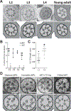
Figure 1.. TZ remodeling in IL2Q cilia
(A) TEM images (upper row) and corresponding tracings (lower row) of IL2Q cilia TZ in cross-section at L2, L3, L4 and young adult (YA) age animals. L2 image is from a hermaphrodite; L3, L4 and YA from males. Scale bar is 100nm. (B) Quantification of the outer dMT and sMTs in IL2Q cilia TZ of L2, L3, L4 and YA males. Each circle (dMTs) and triangle (sMTs) represents measurements from an individual TZ in a different neuron. Errors are SD. The differences between age groups were not significant for either dMT or sMT counts when using the Kruskal-Wallis test with Dunn’s post-hoc correction. See Supplemental Table 1. (C) Comparison of IL2Q TZ lengths in L2 larval and young adult (YA) males. Each symbol represents measurements from an individual TZ in a different neuron. Errors are SD. Asterisk (*) indicates that marked datasets are significantly different at αlpha cutoff set at 0.05 based on pairwise comparison (Mann-Whitney test). (D) TEM images (upper row) and corresponding tracings (lower row) of select cross-sections of larval IL2Q TZs. Arrowheads indicate displaced dMT, A-tubule with hook-like appendage (incomplete dMT), outer sMT without Y link or outer sMT with Y link. Scale bar is 100nm.
Figure 2.
Figure 2.. TZ maturation and axonemal extension in CEM ciliogenesis
(A) TEM images (upper row) and corresponding tracings (lower row) of CEM cilia TZ in cross-section at L2, L3, L4 larvae and young adult (YA) age males. Scale bar is 100nm. (B) Model of growing CEM axoneme anterior of the TZ. Quantity, length and ultrastructure of axonemal MTs is based on serial reconstruction of CEM cilia in L2, L3, L4 and young adult (YA) males. TEM images and corresponding scales show cross-sections of the corresponding CEM axoneme at the focal plane indicated by dashed line. Scale bar is 100nm. (C, D, E) Quantification of the outer dMTs and sMTs (C), TZ lengths (D) and cilia length (E) in CEM cilia of L2, L3, L4 larvae and young adult (YA) males. Each symbol represents measurements from an individual TZ in a different neuron. Errors are SD. Lowercase letters above each age group indicate results of statistical analysis; datasets that do not share a common letter are significantly different at p<0.005 based on Kruskal-Wallis test with Dunn’s post-hoc correction. Also see Supplemental Table 1.
Figure 3.
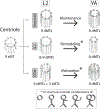
Figure 3.. Summary model of age- and cell-specific TZ structural changes
9-fold symmetry of the centriole templates 9-fold TZ symmetry of sensory cilia in C. elegans larvae. Centriolar structure is depicted as described in (Pelletier et al., 2006). “Amphid” refers to amphid channel cilia whose early ciliogenesis steps are described by Nechipurenko et al., 2017 and Serwas et al., 2017. Inset shows models of axonemal microtubule structures seen in TZ of IL2 and CEM cilia during L2-YA temporal window. YA = young adult.

References

    1. Anand M, and Khanna H (2012). Ciliary transition zone (TZ) proteins RPGR and CEP290: role in photoreceptor cilia and degenerative diseases. Expert Opin. Ther. Targets 16, 541–551. - PMC - PubMed
    1. Avidor-Reiss T, and Leroux MR (2015). Shared and Distinct Mechanisms of Compartmentalized and Cytosolic Ciliogenesis. Curr. Biol. 25, R1143–R1150. - PMC - PubMed
    1. Avidor-Reiss T, Ha A, and Basiri ML (2017). Transition Zone Migration: A Mechanism for Cytoplasmic Ciliogenesis and Postaxonemal Centriole Elongation. Cold Spring Harb Perspect Biol; 9. - PMC - PubMed
    1. Barr MM (2016). Middle Age Has Its Advantages. PLoS Genet. 12, e1006426. - PMC - PubMed
    1. Bruel A-L, Franco B, Duffourd Y, Thevenon J, Jego L, Lopez E, Deleuze J-F, Doummar D, Giles RH, Johnson CA, et al. (2017). Fifteen years of research on Oral–Facial–Digital syndromes: from 1 to 16 causal genes. J. Med. Genet. 54, 371–380. - PMC - PubMed

LinkOut - more resources