Review
. 2019 Jun 1;199(11):1344-1357.
doi: 10.1164/rccm.201807-1212CI.

The Genetics of Pneumothorax

Affiliations
Review

The Genetics of Pneumothorax

Philip M Boone et al. Am J Respir Crit Care Med. .

Abstract

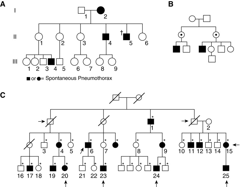
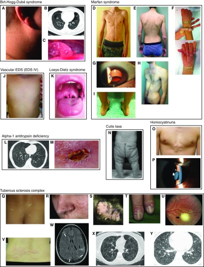
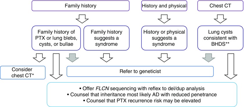
A genetic influence on spontaneous pneumothoraces-those occurring without a traumatic or iatrogenic cause-is supported by several lines of evidence: 1) pneumothorax can cluster in families (i.e., familial spontaneous pneumothorax), 2) mutations in the FLCN gene have been found in both familial and sporadic cases, and 3) pneumothorax is a known complication of several genetic syndromes. Herein, we review known genetic contributions to both sporadic and familial pneumothorax. We summarize the pneumothorax-associated genetic syndromes, including Birt-Hogg-Dubé syndrome, Marfan syndrome, vascular (type IV) Ehlers-Danlos syndrome, alpha-1 antitrypsin deficiency, tuberous sclerosis complex/lymphangioleiomyomatosis, Loeys-Dietz syndrome, cystic fibrosis, homocystinuria, and cutis laxa, among others. At times, pneumothorax is their herald manifestation. These syndromes have serious potential extrapulmonary complications (e.g., malignant renal tumors in Birt-Hogg-Dubé syndrome), and surveillance and/or treatment is available for most disorders; thus, establishing a diagnosis is critical. To facilitate this, we provide an algorithm to guide the clinician in discerning which cases of spontaneous pneumothorax may have a genetic or familial contribution, which cases warrant genetic testing, and which cases should prompt an evaluation by a geneticist.

Keywords: gene; Birt-Hogg-Dubé syndrome; familial spontaneous pneumothorax; genetics; pneumothorax.


Figures

Figure 1.
Figure 1.
Pedigrees demonstrating familial spontaneous pneumothorax. (A) Most pedigrees are consistent with autosomal dominant (AD) inheritance with incomplete penetrance. No causal gene reported for this family. Circles, females; squares, males; solid, pneumothorax; dagger, deceased. Reproduced by permission from Reference . (B) Some pedigrees are also consistent with X-linked recessive inheritance (versus AD with reduced penetrance in females). No causal gene reported for this family. Dot, obligate carrier. Reproduced by permission from Reference . (C) Family with known FLCN mutation. Computed tomography (CT) lung findings (black shading) are more clearly AD than pneumothorax (arrows). Individual 23 has a different bullae phenotype (apical instead of random distribution) and is mutation negative, likely explaining why his mother does not have bullae (different cause of pneumothorax in this branch of family). *CT of the lung performed; diagonal line, deceased. Reproduced by permission from Reference .
Figure 2.
Figure 2.
Physical examination findings of pneumothorax-associated syndromes. (AC) Birt-Hogg-Dubé syndrome. (A) Fibrofolliculomas of the neck (159). (B) Lung cysts and bullae; extra-apical location is characteristic (159). (C) Pleural blebs on the surface of the left lower lobe; these can be missed on computed tomography but seen during thoracoscopy/video-assisted thorascopic surgery (159). (DJ) Marfan syndrome. (D) Marfanoid body habitus (90). (E) Scoliosis, striae, reduced elbow extension (160). (F) Positive thumb and wrist signs indicating arachnodactyly (160). (G) Lens dislocation (161). (H) Pectus excavatum (160). (I) Hindfoot deformity (160). (J) Vascular (type IV) Ehlers-Danlos syndrome: translucent skin on the torso of an infant (162). (K) Loeys-Dietz syndrome: bifid uvula (163) (L and M) Alpha-1 antitrypsin deficiency. (L) Panlobular emphysema (case courtesy of Dr. Jeremy Jones, Radiopaedia.org, rID:13441). (M) Panniculitis (164). (N) Cutis laxa: autosomal recessive cutis laxa IIa (165). (O and P) Homocystinuria. (O) Chest wall deformity (166). (P) Lens dislocation (167). (QY) Tuberous sclerosis complex. (Q) Hypopigmented macules (ash leaf spots) (168). (R) Angiofibromas (168). (S) Fibrous plaques (168). (T) Ungual fibromas (168). (U) Retinal hamartoma (168). (V) Shagreen patch (168). (W) Cortical dysplasia (tubers, white arrows; radial migration lines, black arrow) (168). (X) Lymphangioleiomyomatosis (168). (Y) Multifocal micronodular pneumocyte hyperplasia (169). Images are reproduced by permission from the sources cited above.
Figure 3.
Figure 3.
Proposed algorithm for identifying spontaneous pneumothoraces with a genetic basis. Algorithm should be applied to all patients with spontaneous pneumothorax. Gray arrows are optional, based on the practitioner’s comfort. *FLCN testing in cases of absent family history is still reasonable, so long as a chest computed tomography (CT) scan, if previously obtained, is not inconsistent with Birt-Hogg-Dubé syndrome (BHDS). **Chest CT findings in BHDS include bilateral, multifocal, predominately lower-lobe cysts. AD = autosomal dominant; PTX = pneumothorax.
Figure 4.
Figure 4.
History, physical examination, and imaging findings that raise the possibility of a syndromic cause of pneumothorax. The major features (x-axis) of syndromes predisposing to pneumothorax (y-axis) are shown. Personal history/family history/physical examination findings are in blue. Imaging findings are in red. The skin findings of Birt-Hogg-Dubé syndrome (BHDS) are age dependent; thus, skin examinations of the parents or even grandparents may at times be more informative than for the proband. A1ATD = alpha-1 antitrypsin deficiency; CF = cystic fibrosis; LDS = Loeys-Dietz syndrome; TSC-LAM = lymphangioleiomyomatosis in an individual with tuberous sclerosis; vEDS = vascular Ehlers-Danlos syndrome.

References

    1. Bense L, Lewander R, Eklund G, Hedenstierna G, Wiman LG. Nonsmoking, non-alpha 1-antitrypsin deficiency-induced emphysema in nonsmokers with healed spontaneous pneumothorax, identified by computed tomography of the lungs. Chest. 1993;103:433–438. - PubMed
    1. Withers JN, Fishback ME, Kiehl PV, Hannon JL. Spontaneous pneumothorax: suggested etiology and comparison of treatment methods. Am J Surg. 1964;108:772–776. - PubMed
    1. Bense L, Eklund G, Wiman LG. Smoking and the increased risk of contracting spontaneous pneumothorax. Chest. 1987;92:1009–1012. - PubMed
    1. Abolnik IZ, Lossos IS, Zlotogora J, Brauer R. On the inheritance of primary spontaneous pneumothorax. Am J Med Genet. 1991;40:155–158. - PubMed
    1. Ren HZ, Zhu CC, Yang C, Chen SL, Xie J, Hou YY, et al. Mutation analysis of the FLCN gene in Chinese patients with sporadic and familial isolated primary spontaneous pneumothorax. Clin Genet. 2008;74:178–183. - PubMed

Publication types