

GSK923295 as a potential antihepatocellular carcinoma agent causing delay on liver regeneration after partial hepatectomy
- PMID: 30681497
- PMCID: PMC6595801
- DOI: 10.1097/CM9.0000000000000053
GSK923295 as a potential antihepatocellular carcinoma agent causing delay on liver regeneration after partial hepatectomy
Abstract
Background: The clinical trials emerged centromere protein E inhibitor GSK923295 as a promising anticancer drug, but its function in hepatocellular carcinoma (HCC) remain needs to be fully elucidated, especially as chemotherapy after hepatectomy for liver tumors. We aimed to describe anti-HCC activities of GSK923295 and compare its antiproliferative effects on liver regeneration after partial hepatectomy (PH).
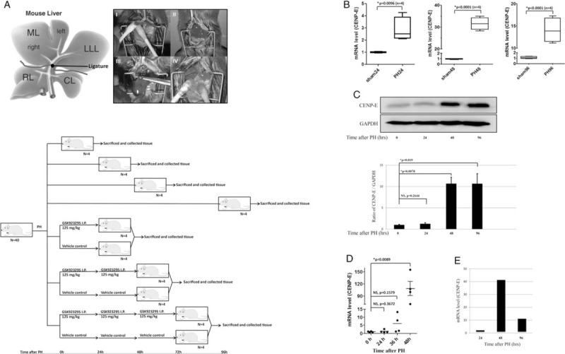
Methods: All subjects were randomized to treatment with either vehicle or GSK923295. Antitumor activity of GSK923295 was assessed by xenograft growth assays. The C57BL/6 mice were subjected to 70% PH and the proliferation was calculated by liver coefficient, further confirmed by immunohistochemistry. The proliferation and cell cycle analysis of liver cell AML12 and HCC cells LM3, HUH7, and HepG2 were investigated using the cell counting kit-8 assay and Flow Cytometry. The chromosome misalignment and segregation in AML12 cells were visualized by immunofluorescence.
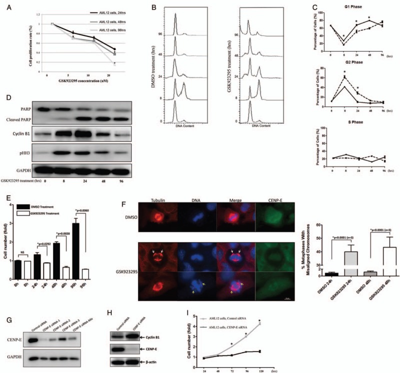
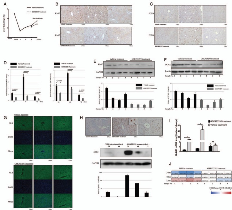
Results: Treatment with GSK923295 induced antiproliferation in HCC cell lines. It also caused delay on HCC tumor growth instead of regression both in a HCC cell line xenograft model and patient-derived tumor xenograft model. With microarray analysis, CENtromere Protein E was gradually increased in mouse liver after PH. Exposure of liver cells to GSK923295 resulted in delay on a cell cycle in mitosis with a phenotype of misaligned chromosomes and chromosomes clustered. In 70% PH mouse model, GSK923295 treatment also remarkably reduced liver regeneration in later stage, in parallel with the mitotic marker phospho-histone H3 elevation.
Conclusion: The anticancer drug GSK923295 causes a significant delay on HCC tumor growth and liver regeneration after PH in later stage.
Figures

Similar articles
-
First-time-in-human study of GSK923295, a novel antimitotic inhibitor of centromere-associated protein E (CENP-E), in patients with refractory cancer.Cancer Chemother Pharmacol. 2012 Mar;69(3):733-41. doi: 10.1007/s00280-011-1756-z. Epub 2011 Oct 22. Cancer Chemother Pharmacol. 2012. PMID: 22020315 Clinical Trial.
-
Mitogen-activated protein kinase (MEK/ERK) inhibition sensitizes cancer cells to centromere-associated protein E inhibition.Int J Cancer. 2013 Feb 1;132(3):E149-57. doi: 10.1002/ijc.27781. Epub 2012 Sep 28. Int J Cancer. 2013. PMID: 22948716 Free PMC article.
-
Pgp efflux pump decreases the cytostatic effect of CENP-E inhibitor GSK923295.Cancer Lett. 2015 May 28;361(1):97-103. doi: 10.1016/j.canlet.2015.02.040. Epub 2015 Feb 25. Cancer Lett. 2015. PMID: 25725449
-
Antitumor effect of vascular endothelial growth factor inhibitor sunitinib in preclinical models of hepatocellular carcinoma.Eur J Gastroenterol Hepatol. 2012 May;24(5):563-74. doi: 10.1097/MEG.0b013e328350916f. Eur J Gastroenterol Hepatol. 2012. PMID: 22314934
-
Antitumor Effects of Quercetin in Hepatocarcinoma In Vitro and In Vivo Models: A Systematic Review.Nutrients. 2019 Nov 25;11(12):2875. doi: 10.3390/nu11122875. Nutrients. 2019. PMID: 31775362 Free PMC article.
Cited by
-
Kinesin-7 CENP-E in tumorigenesis: Chromosome instability, spindle assembly checkpoint, and applications.Front Mol Biosci. 2024 Mar 15;11:1366113. doi: 10.3389/fmolb.2024.1366113. eCollection 2024. Front Mol Biosci. 2024. PMID: 38560520 Free PMC article. Review.
-
Recent updates of centromere proteins in hepatocellular carcinoma: a review.Infect Agent Cancer. 2025 Feb 6;20(1):7. doi: 10.1186/s13027-024-00630-2. Infect Agent Cancer. 2025. PMID: 39915786 Free PMC article. Review.
-
Generation and application of patient-derived xenograft models in pancreatic cancer research.Chin Med J (Engl). 2019 Nov 20;132(22):2729-2736. doi: 10.1097/CM9.0000000000000524. Chin Med J (Engl). 2019. PMID: 31725451 Free PMC article. Review.
-
The role of kinesin superfamily proteins in hepatocellular carcinoma.Med Oncol. 2024 Oct 14;41(11):271. doi: 10.1007/s12032-024-02497-0. Med Oncol. 2024. PMID: 39400594 Review.
-
The role of kinesin family members in hepatobiliary carcinomas: from bench to bedside.Biomark Res. 2024 Mar 3;12(1):30. doi: 10.1186/s40364-024-00559-z. Biomark Res. 2024. PMID: 38433242 Free PMC article. Review.
References
-
- Torre LA, Bray F, Siegel RL, Ferlay J, Lortet-Tieulent J, Jemal A. Global cancer statistics, 2012. CA Cancer J Clin 2015; 65:87–108. doi: 10.3322/caac.21262. - PubMed
-
- Forner A, Llovet JM, Bruix J. Hepatocellular carcinoma. Lancet 2012; 379:1245–1255. doi: 10.1016/S0140-6736(11)61347-0. - PubMed
-
- Sapisochin G, Goldaracena N, Laurence JM, Dib M, Barbas A, Ghanekar A, et al. The extended Toronto criteria for liver transplantation in patients with hepatocellular carcinoma: a prospective validation study. Hepatology 2016; 64:2077–2088. doi: 10.1002/hep.28643. - PubMed
-
- Ban D, Shimada K, Yamamoto Y, Nara S, Esaki M, Sakamoto Y, et al. Efficacy of a hepatectomy and a tumor thrombectomy for hepatocellular carcinoma with tumor thrombus extending to the main portal vein. J Gastrointest Surg 2009; 13:1921–1928. doi: 10.1007/s11605-009-0998-0. - PubMed
-
- Weese JL, Ottery FD, Emoto SE. Do operations facilitate tumor growth? An experimental model in rats. Surgery 1986; 100:273–277. - PubMed
MeSH terms
Substances
LinkOut - more resources
Full Text Sources
Medical
