Optical coherence tomography angiography in unilateral multifocal choroiditis and panuveitis: A case report
- PMID: 30681623
- PMCID: PMC6358364
- DOI: 10.1097/MD.0000000000014259
Optical coherence tomography angiography in unilateral multifocal choroiditis and panuveitis: A case report
Abstract
Rationale: Optical coherence tomography angiography (OCT-A) has the advantage to visualize the microvascular structure of the retina in vivo and was utilized clinically in various neovascular retinal diseases. The OCT-A has also been used to examine the lesion in multifocal choroiditis and panuveitis (MCP). This study aimed to describe a case of MCP and present the disease process of a punched-out lesion in the chorioretina with neovascular activity using OCT-A.
Patients concerns: A 32-year-old female Caucasian patient presented with a 2-week history of progressive blurred vision in her right eye with photophobia and a diminished temporal visual field. On presentation, her best corrected visual acuity was 6/60 in the right eye with a prominent anterior uveitis seen under slit lamp examination.
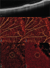
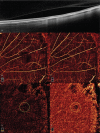
Diagnoses: Dilated fundus examination of the right eye showed vitritis and multiple, punched-out yellowish-white lesions over the peripheral retina. Additional multimodal imaging (MMI) were done including fluorescein angiography (FA), indocyanine green angiography (ICGA) and fundus autofluorescence (FAF), which all revealed characteristic findings of MCP. In general, the diagnosis of unilateral MCP was made. Furthermore, one of the punched-out lesions in the right eye was particularly selected and examined under OCT and OCT-A, which revealed a subretinal elevated lesion with high flow signal under OCT-A.
Interventions: Treatment with oral prednisolone at 30 mg daily with topical prednisolone acetate 1% every 2 hours were prescribed, which were gradually tapered down within a 2-month course.
Outcomes: The patient's best corrected visual acuity of the right eye returned to 6/6 at 2 months after the diagnosis. The flow signal in the OCT-A study of the punched-out lesion had also resolved after steroid treatment.
Lessons: The MCP is an uncommon uveitis with multiple inflammatory chorioretinal lesions. Using multimodal imaging technique, physicians can better differentiate these lesions for diagnosis and for further monitoring. Our results demonstrated that these chorioretinal lesions in MCP may display neovascular activities that might not be seen easily using conventional FA or ICGA study. With OCT-A, ophthalmologists could identify and monitor subtle choroidal neovascularization (CNV) changes over these punched-out lesions.
Conflict of interest statement
The authors have no conflicts of interest to disclose.
Figures


References
-
- Quillen DA, Davis JB, Gottlieb JL, et al. The white dot syndromes. Am J Ophthalmol 2004;137:538–50. doi:10.1016/j.ajo.2004.01.053. - PubMed
-
- Dreyer RF, Gass JDM. Multifocal choroiditis and panuveitis: a syndrome that mimics ocular histoplasmosis. Arch Ophthalmol 1984;102:1776–84. doi:10.1001/archopht.1984.01040031440019. - PubMed
Publication types
MeSH terms
LinkOut - more resources
Full Text Sources
Miscellaneous

