

Protein Methyltransferase Inhibition Decreases Endocrine Specification Through the Upregulation of Aldh1b1 Expression
- PMID: 30681750
- PMCID: PMC6850398
- DOI: 10.1002/stem.2979
Protein Methyltransferase Inhibition Decreases Endocrine Specification Through the Upregulation of Aldh1b1 Expression
Abstract
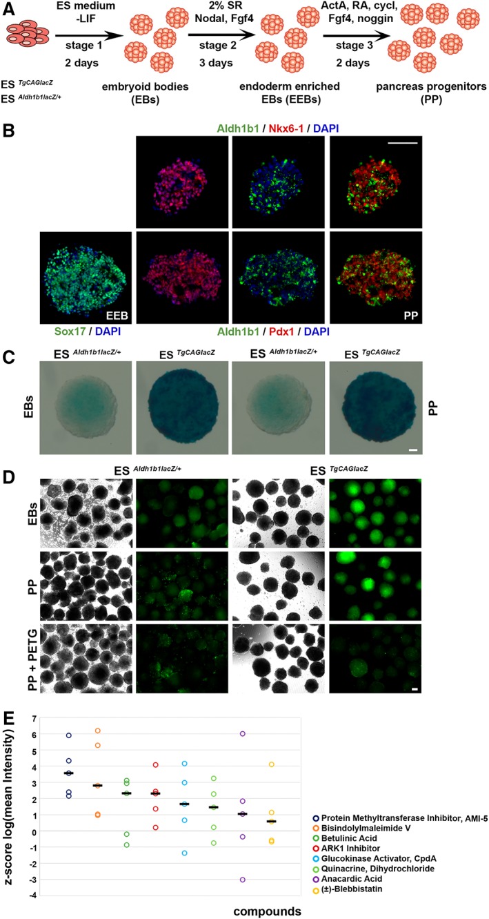
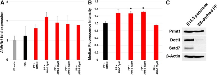
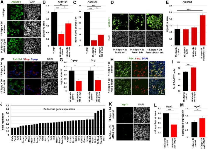
Understanding the mechanisms that promote the specification of pancreas progenitors and regulate their self-renewal and differentiation will help to maintain and expand pancreas progenitor cells derived from human pluripotent stem (hPS) cells. This will improve the efficiency of current differentiation protocols of hPS cells into β-cells and bring such cells closer to clinical applications for the therapy of diabetes. Aldehyde dehydrogenase 1b1 (Aldh1b1) is a mitochondrial enzyme expressed specifically in progenitor cells during mouse pancreas development, and we have shown that its functional inactivation leads to accelerated differentiation and deficient β-cells. In this report, we aimed to identify small molecule inducers of Aldh1b1 expression taking advantage of a mouse embryonic stem (mES) cell Aldh1b1 lacZ reporter line and a pancreas differentiation protocol directing mES cells into pancreatic progenitors. We identified AMI-5, a protein methyltransferase inhibitor, as an Aldh1b1 inducer and showed that it can maintain Aldh1b1 expression in embryonic pancreas explants. This led to a selective reduction in endocrine specification. This effect was due to a downregulation of Ngn3, and it was mediated through Aldh1b1 since the effect was abolished in Aldh1b1 null pancreata. The findings implicated methyltransferase activity in the regulation of endocrine differentiation and showed that methyltransferases can act through specific regulators during pancreas differentiation. Stem Cells 2019;37:640-651.
Keywords: Aldehyde dehydrogenase 1b1; Embryonic stem cell pancreas differentiation; Endocrine specification; Pancreas development; Pancreatic progenitors; β-Cells.
© 2019 The Authors. Stem Cells published by Wiley Periodicals, Inc. on behalf of AlphaMed Press 2019.
Conflict of interest statement
The authors indicated no potential conflicts of interest.
Figures

Similar articles
-
Aldh1b1 expression defines progenitor cells in the adult pancreas and is required for Kras-induced pancreatic cancer.Proc Natl Acad Sci U S A. 2019 Oct 8;116(41):20679-20688. doi: 10.1073/pnas.1901075116. Epub 2019 Sep 23. Proc Natl Acad Sci U S A. 2019. PMID: 31548432 Free PMC article.
-
Aldehyde dehydrogenase activity is necessary for beta cell development and functionality in mice.Diabetologia. 2016 Jan;59(1):139-150. doi: 10.1007/s00125-015-3784-4. Epub 2015 Oct 31. Diabetologia. 2016. PMID: 26518685 Free PMC article.
-
ALDH1B1 is a potential stem/progenitor marker for multiple pancreas progenitor pools.Dev Biol. 2013 Feb 1;374(1):153-63. doi: 10.1016/j.ydbio.2012.10.030. Epub 2012 Nov 6. Dev Biol. 2013. PMID: 23142317 Free PMC article.
-
PDX1, Neurogenin-3, and MAFA: critical transcription regulators for beta cell development and regeneration.Stem Cell Res Ther. 2017 Nov 2;8(1):240. doi: 10.1186/s13287-017-0694-z. Stem Cell Res Ther. 2017. PMID: 29096722 Free PMC article. Review.
-
Revisiting the immunocytochemical detection of Neurogenin 3 expression in mouse and man.Diabetes Obes Metab. 2016 Sep;18 Suppl 1:10-22. doi: 10.1111/dom.12718. Diabetes Obes Metab. 2016. PMID: 27615127 Review.
Cited by
-
Aldh1b1 expression defines progenitor cells in the adult pancreas and is required for Kras-induced pancreatic cancer.Proc Natl Acad Sci U S A. 2019 Oct 8;116(41):20679-20688. doi: 10.1073/pnas.1901075116. Epub 2019 Sep 23. Proc Natl Acad Sci U S A. 2019. PMID: 31548432 Free PMC article.
-
Generation and application of novel hES cell reporter lines for the differentiation and maturation of hPS cell-derived islet-like clusters.Sci Rep. 2024 Aug 27;14(1):19863. doi: 10.1038/s41598-024-69645-4. Sci Rep. 2024. PMID: 39191834 Free PMC article.
References
-
- Rezania A, Bruin JE, Arora P et al. Reversal of diabetes with insulin‐producing cells derived in vitro from human pluripotent stem cells. Nat Biotechnol 2014;32:1121–1133. - PubMed
-
- Kawaguchi Y, Cooper B, Gannon M et al. The role of the transcriptional regulator Ptf1a in converting intestinal to pancreatic progenitors. Nat Genet 2002;32:128–134. - PubMed
Publication types
MeSH terms
Substances
LinkOut - more resources
Full Text Sources
Other Literature Sources
Medical
Molecular Biology Databases
Miscellaneous
