. 2019 Aug 6;35(31):10061-10067.
doi: 10.1021/acs.langmuir.8b03692. Epub 2019 Feb 8.

Fibrin-Targeted Polymerized Shell Microbubbles as Potential Theranostic Agents for Surgical Adhesions

Affiliations

Fibrin-Targeted Polymerized Shell Microbubbles as Potential Theranostic Agents for Surgical Adhesions

Catherine A Gormley et al. Langmuir. .

Abstract

The development of new therapies for surgical adhesions has proven to be difficult as there is no consistently effective way to assess treatment efficacy in clinical trials without performing a second surgery, which can result in additional adhesions. We have developed lipid microbubble formulations that use a short peptide sequence, CREKA, to target fibrin, the molecule that forms nascent adhesions. These targeted polymerized shell microbubbles (PSMs) are designed to allow ultrasound imaging of early adhesions for diagnostic purposes and for evaluating the success of potential treatments in clinical trials while acting as a possible treatment. In this study, we show that CREKA-targeted microbubbles preferentially bind fibrin over fibrinogen and are stable for long periods of time (∼48 h), that these bound microbubbles can be visualized by ultrasound, and that neither these lipid-based bubbles nor their diagnostic-ultrasound-induced vibrations damage mesothelial cells in vitro. Moreover, these bubbles show the potential to identify adhesionlike fibrin formations and may hold promise in blocking or breaking up fibrin formations in vivo.


Conflict of interest statement

The authors declare no competing financial interest.

Figures
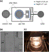
Figure 1.
Figure 1.
(a) Schematic drawing of the microfluidic stepemulsification geometry used to produce microbubbles. The chip had channels with different heights of 1 μm (blue) and 20 μm (gray). (b) Optical image of the microchannel array at the end of the microfluidic chip containing 5-μm-wide channels for bubble production (scale bar 100 μm). (c) Image of the microfluidic chip (scale bar 1 cm).
Figure 2.
Figure 2.
The polymerized shell microbubble (PSM) stability and diameter do not change significantly over time. The mean diameters of nontargeted (NT-PSM) and fibrin-targeted (T-PSM) polymerized shell microbubbles (PSMs) are shown over time. The PSMs were incubated in PBS or media at 37 °C. The data are expressed as the mean ± SD.
Figure 3.
Figure 3.
LP-9 cellular viability after PSM exposure. LP-9 cells were exposed to 105 and 107 fibrin-targeted and nontargeted PSMs (T-PSM and NT-PSM, respectively) for 2, 24, and 48 h prior to assessing the cellular viability. Viability is expressed as the total number of dead cells divided by the total number of cells (n = 3). Fluorescent images were obtained for each time point and condition and were analyzed and quantified using ImageJ. Data is expressed as the mean ± SEM.
Figure 4.
Figure 4.
LP-9 cellular viability after treatment with PSM and ultrasound. LP-9 cells were exposed to either 105 targeted or nontargeted PSM (T-PSM and NT-PSM, respectively) in the absence and presence of ultrasound treatment. Cellular viability was assessed in triplicate experiments using the LIVE/DEAD assay. Data is expressed as the mean ± SEM.
Figure 5.
Figure 5.
Assessment of the specificity of fibrin binding of targeted PSMs. (a) Targeted microbubbles bind specifically to fibrin. Agarose solution (1%) was added and allowed to solidify in 100 cm Petri dishes. Fibrinogen, fibrin, or no addition (control) was added to the agarose layers. Fibrin-targeted or nontargeted PSMs (105, T-PSM and NT-PSM, respectively) were added to each plate and incubated for 30 min at room temperature. Unbound PSMs were rinsed, and the fluorescence (ex/em 547/580) for each condition was measured using an OptiMax plate reader. Data is expressed as the mean ± SEM. (b) Targeted microbubbles (T-PSM) bound to fibrin are visible in B-mode ultrasound. Agarose gels (1%) were prepared in 100 cm Petri dishes with fibrin blotted (with “no fibrin” controls). Plates were exposed to nontargeted or fibrin-targeted PSMs (T-PSM, NT-PSM) for 30 min and then washed. B-mode ultrasound images were taken, and ImageJ was used to calculate grayscale values for fibrin and nonfibrin regions or similarly located regions on the “no fibrin” (− fibrin) plates. These values were averaged, and the fibrin (or corresponding) region’s average was divided by the “no fibrin” region. Error bars are uncertainties in the ratio.

References

    1. Centers for Disease Control. Number of All-Listed Procedures for Discharges from Short-Stay Hospitals, by Procedure Category and Age: United States; 2010.
    1. Schnüriger B; Barmparas G; Branco BC; Lustenberger T; Inaba K; Demetriades D Prevention of Postoperative Peritoneal Adhesions: A Review of the Literature. Am. J. Surg 2011, 201 (1), 111–121. - PubMed
    1. Arung W; Meurisse M; Detry O Pathophysiology and Prevention of Postoperative Peritoneal Adhesions. World J. Gastroenterol. WJG 2011, 17 (41), 4545–4553. - PMC - PubMed
    1. Lyell DJ; Caughey AB; Hu E; Daniels K Peritoneal Closure at Primary Cesarean Delivery and Adhesions. Obstet. Gynecol 2005, 106 (2), 275–280. - PubMed
    1. Duron J-J Postoperative Intraperitoneal Adhesion Pathophysiology. Colorectal Dis. Off. J. Assoc. Coloproctology G. B. Irel 2007, 9 (Suppl 2), 14–24. - PubMed

Publication types

MeSH terms

Substances