. 2019 Apr;160(4):824-832.
doi: 10.1097/j.pain.0000000000001458.

Kappa opioid signaling in the central nucleus of the amygdala promotes disinhibition and aversiveness of chronic neuropathic pain

Affiliations

Kappa opioid signaling in the central nucleus of the amygdala promotes disinhibition and aversiveness of chronic neuropathic pain

Edita Navratilova et al. Pain. 2019 Apr.

Abstract

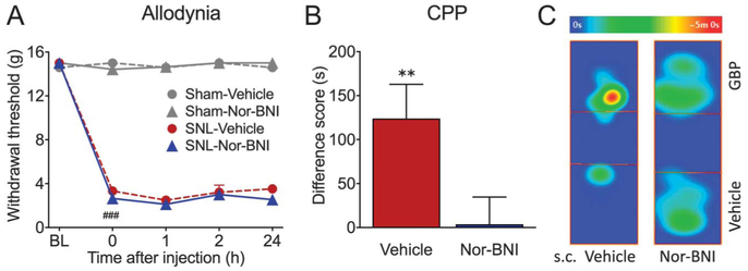
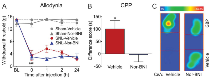
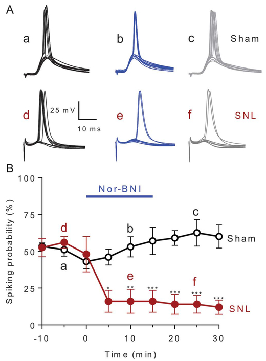
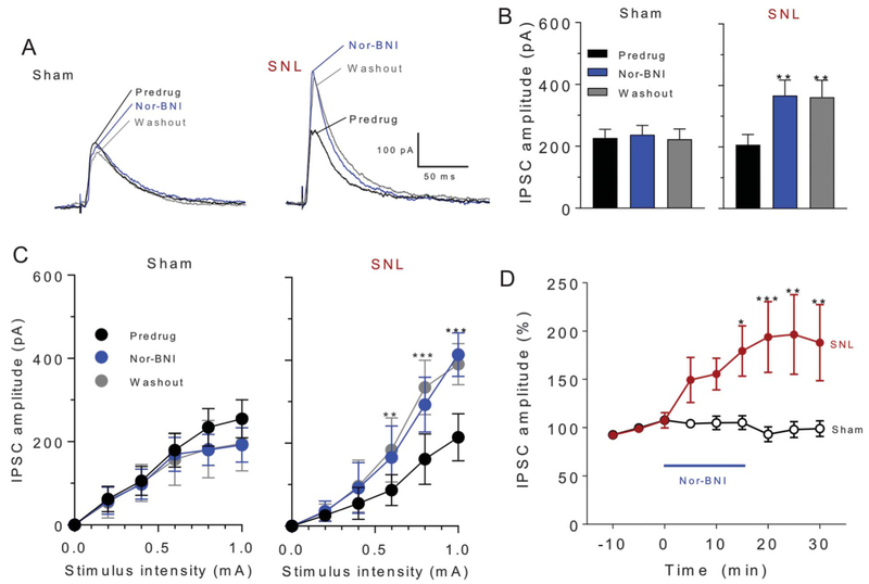
Chronic pain is associated with neuroplastic changes in the amygdala that may promote hyper-responsiveness to mechanical and thermal stimuli (allodynia and hyperalgesia) and/or enhance emotional and affective consequences of pain. Stress promotes dynorphin-mediated signaling at the kappa opioid receptor (KOR) in the amygdala and mechanical hypersensitivity in rodent models of functional pain. Here, we tested the hypothesis that KOR circuits in the central nucleus of the amygdala (CeA) undergo neuroplasticity in chronic neuropathic pain resulting in increased sensory and affective pain responses. After spinal nerve ligation (SNL) injury in rats, pretreatment with a long-acting KOR antagonist, nor-binaltorphimine (nor-BNI), subcutaneously or through microinjection into the right CeA, prevented conditioned place preference (CPP) to intravenous gabapentin, suggesting that nor-BNI eliminated the aversiveness of ongoing pain. By contrast, systemic or intra-CeA administration of nor-BNI had no effect on tactile allodynia in SNL animals. Using whole-cell patch-clamp electrophysiology, we found that nor-BNI decreased synaptically evoked spiking of CeA neurons in brain slices from SNL but not sham rats. This effect was mediated through increased inhibitory postsynaptic currents, suggesting tonic disinhibition of CeA output neurons due to increased KOR activity as a possible mechanism promoting ongoing aversive aspects of neuropathic pain. Interestingly, this mechanism is not involved in SNL-induced mechanical allodynia. Kappa opioid receptor antagonists may therefore represent novel therapies for neuropathic pain by targeting aversive aspects of ongoing pain while preserving protective functions of acute pain.


Conflict of interest statement

Conflict of interest statement

The authors have no conflict of interest to declare.

Figures

Figure 1.
Figure 1.
Systemic nor-BNI relieves aversiveness of ongoing pain but not tactile allodynia in SNL rats. (A) After SNL surgery, rats develop long-lasting mechanical allodynia in the injured paw, demonstrated as reduced tactile paw withdrawal thresholds. Systemic administration of nor-BNI (3 mg/kg, s.c.) or vehicle (saline, s.c.) had no effect on tactile allodynia in either sham or SNL animals during a 24-hour time course. N = 4–8; ###P < 0.001. (B) SNL animals were pretreated with either nor-BNI (3 mg/kg, s.c.) or vehicle (saline, s.c.) 1 day before CPP conditioning. In vehicle pretreated rats, administration of gabapentin (50 mg/kg, i.v.) produced significant place preference for the GBP-paired chamber, shown as a positive difference score, suggesting the presence of ongoing pain that is relieved by gabapentin treatment. N = 16, **P = 0.006. By contrast, nor-BNI pretreated animals show no chamber preference, indicating that nor-BNI relieved ongoing pain. N = 13. (C) Representative heat maps demonstrating time spent in different locations in the CPP box on test day. SNL rat pretreated with subcutaneous (s.c.) saline spent more time in the GBP-paired chamber, whereas nor-BNI pretreated rat spent equivalent time in both chambers. Data are expressed as mean ± SEM. CPP, conditioned place preference; nor-BNI, nor-binaltorphimine; SNL, spinal nerve ligation.
Figure 2.
Figure 2.
Blockade of KOR in the CeA relieves aversiveness of ongoing neuropathic pain in SNL rats. (A) SNL rats displayed significantly reduced paw withdrawal thresholds, indicating presence of tactile allodynia. Microinjection of nor-BNI (2.5 μg/0.5 μL) or vehicle (saline, 0.5 μL) into the right CeA had no effect on tactile allodynia in either sham or SNL animals during a 24-hour time course. N = 3–5; ###P < 0.001. (B) Gabapentin (50 mg/kg; i.v.) produces CPP in SNL rats pretreated 24 hours before conditioning with vehicle into the right CeA (N = 13, *P = 0.011), but not in SNL rats microinjected with nor-BNI (2.5 μg/0.5 μL) (N = 18). (C) Representative heat maps demonstrating time spent in different locations in the CPP box on test day. SNL rat pretreated with saline in the CeA spent more time in the GBP-paired chamber on test day. By contrast, CeA nor-BNI pretreated rat spent equivalent time in both chambers. Data are expressed as mean ± SEM. CPP, conditioned place preference; KOR, kappa opioid receptor; nor-BNI, nor-binaltorphimine; SNL, spinal nerve ligation.
Figure 3.
Figure 3.
Synaptically evoked spiking. Nor-BNI (1 μM, 15 minutes) decreased synaptically evoked spiking (E-S coupling) in neurons in the laterocapsular division of the central nucleus of the amygdala (CeLC) in brain slices from SNL but not sham rats, 4 weeks after surgery. (A) Current-clamp recordings of action potentials (spikes) evoked by electrical stimulation of the dorsomedial fiber tract into CeLC. Voltage traces (a–f) show responses of 1 neuron recorded at −60 mV (a and d) before, (b and e) during, and (c and f) after washout of nor-BNI in slices from sham or SNL rats. Each trace is the response to one synaptic stimulation. (B) Time course of drug effects. Graph shows averaged values (±SEM) for the sample of neurons from sham (n = 10 neurons) and SNL rats (n = 5 neurons). Each symbol is the mean value for all neurons in each sample. In each neuron, a series of 10 synaptically evoked responses was measured per time point to determine the spiking probability. Bars represent SEM. Two-way ANOVA with Bonferroni post hoc test’s comparison between sham and SNL groups (*P < 0.05, **P < 0.01, ***P < 0.001; F8,110 = 4.926). ANOVA, analysis of variance; nor-BNI, norbinaltorphimine; SNL, spinal nerve ligation.
Figure 4.
Figure 4.
Synaptic inhibition. Nor-BNI (1 μM) increased inhibitory synaptic currents (IPSCs) in neurons in the CeLC in brain slices from SNL rats but not sham controls, 4 weeks after surgery. Electrophysiological recordings were performed from regular spiking neurons after electrical stimulation of the dorsomedial fiber tract. (A) Current traces from an individual CeLC neuron from a sham rat and another neuron from an SNL rat show voltage-clamp recordings of IPSCs (at 0 mV) evoked by electrical synaptic stimulation (600 μA) before and during drug application and during washout. (B) Mean IPSC amplitudes (±SEM) evoked at 600 μA in the sample of neurons from sham and SNL rats (sham, n = 9 neurons, F2,23 = 0.060, P = 0.942; SNL, n = 7 neurons, F2,14 = 10.98, *P < 0.01; repeated-measures ANOVA with Tukey’s post hoc tests). Each symbol is the mean value for all neurons in each sample. In each neuron, a series of 3 synaptically evoked responses was measured per time point. (C) Input–output function of IPSCs in brain slices from sham and SNL rats shows significant effects of nor-BNI in the neuropathic pain model but not in sham control conditions (sham, n = 5 neurons, F2,10 = 0.725, P = 0.488; SNL, n = 6 neurons, F2,10 = 13.95, **, *** P < 0.01, 0.001, repeated-measures 2-way ANOVA with Tukey’s post hoc tests). (D) Time course of drug effects on IPSCs evoked at 600 μA. Nor-BNI increased IPSCs in CeLC neurons from SNL rats (n = 7 neurons) but not sham rats (n = 9 neurons). Graph shows averaged values (±SEM). F8,130 = 2.384, *P < 0.05, ** P < 0.01, ***P < 0.001; 2-way ANOVA with Tukey’s post hoc tests. ANOVA, analysis of variance; nor-BNI, nor-binaltorphimine; SNL, spinal nerve ligation.

Similar articles

Cited by

References

    1. Arora V, Martin TJ, Aschenbrenner CA, Hayashida K, Kim SA, Parker RA, Eisenach JC, Peters CM. Psychosocial stress delays recovery of postoperative pain following incisional surgery in the rat. Neuroscience 2018;382:35–47. - PMC - PubMed
    1. Baliki MN, Apkarian AV. Nociception, pain, negative moods, and behavior selection. Neuron 2015;87:474–91. - PMC - PubMed
    1. Bannister K, Qu C, Navratilova E, Oyarzo J, Xie JY, King T, Dickenson AH, Porreca F. Multiple sites and actions of gabapentin-induced relief of ongoing experimental neuropathic pain. PAIN 2017;158:2386–95. - PMC - PubMed
    1. Beckerman MA, Van Kempen TA, Justice NJ, Milner TA, Glass MJ. Corticotropin-releasing factor in the mouse central nucleus of the amygdala: ultrastructural distribution in NMDA-NR1 receptor subunit expressing neurons as well as projection neurons to the bed nucleus of the stria terminalis. Exp Neurol 2013;239:120–32. - PMC - PubMed
    1. Borsook D, Maleki N, Becerra L, McEwen B. Understanding migraine through the lens of maladaptive stress responses: a model disease of allostatic load. Neuron 2012;73:219–34. - PubMed

MeSH terms