. 2019 Jan 25;15(1):e1007560.
doi: 10.1371/journal.ppat.1007560. eCollection 2019 Jan.

Influenza-induced immune suppression to methicillin-resistant Staphylococcus aureus is mediated by TLR9

Affiliations

Influenza-induced immune suppression to methicillin-resistant Staphylococcus aureus is mediated by TLR9

Giovanny J Martínez-Colón et al. PLoS Pathog. .

Abstract

Bacterial lung infections, particularly with methicillin-resistant Staphylococcus aureus (MRSA), increase mortality following influenza infection, but the mechanisms remain unclear. Here we show that expression of TLR9, a microbial DNA sensor, is increased in murine lung macrophages, dendritic cells, CD8+ T cells and epithelial cells post-influenza infection. TLR9-/- mice did not show differences in handling influenza nor MRSA infection alone. However, TLR9-/- mice have improved survival and bacterial clearance in the lung post-influenza and MRSA dual infection, with no difference in viral load during dual infection. We demonstrate that TLR9 is upregulated on macrophages even when they are not themselves infected, suggesting that TLR9 upregulation is related to soluble mediators. We rule out a role for elevations in interferon-γ (IFNγ) in mediating the beneficial MRSA clearance in TLR9-/- mice. While macrophages from WT and TLR9-/- mice show similar phagocytosis and bacterial killing to MRSA alone, following influenza infection, there is a marked upregulation of scavenger receptor A and MRSA phagocytosis as well as inducible nitric oxide synthase (Inos) and improved bacterial killing that is specific to TLR9-deficient cells. Bone marrow transplant chimera experiments and in vitro experiments using TLR9 antagonists suggest TLR9 expression on non-hematopoietic cells, rather than the macrophages themselves, is important for regulating myeloid cell function. Interestingly, improved bacterial clearance post-dual infection was restricted to MRSA, as there was no difference in the clearance of Streptococcus pneumoniae. Taken together these data show a surprising inhibitory role for TLR9 signaling in mediating clearance of MRSA that manifests following influenza infection.


Conflict of interest statement

The authors have declared that no competing interests exist.

Figures
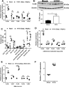
Fig 1
Fig 1. TLR9 overexpression in lung immune cells post-IAV infection.
(A) Relative gene expression of TLRs (9, 7, 3, 2, 4) by RTqPCR and (B) western blotting of TLR9 and β-actin. RNA and protein were isolated from lung leukocytes post-collagenase digestion in mice infected with 100 PFUs of H1N1 (PR8) for 5 days or placebo (PBS). β-actin was used to normalize RNA in samples. (C) Frequency of TLR9+ cells measured by flow cytometry in lung immune cells post-collagenase digestion. Gating was as follows: CD4+ T cells (CD45+,CD90.2+, CD3+, CD4+), CD8+ T cells (CD45+,CD90.2+, CD3+, CD4-, CD8+), natural killer cells (CD45+,CD90.2+, NKp46+), B cells (CD45+,CD90.2-, CD19+), neutrophils (CD45+, CD11b+, LY6G+), interstitial macrophages (CD45+, CD64+, CD11b+, F4/80+), alveolar macrophages (CD45+, CD64+, CD11C+, Siglec F+) and dendritic cells (CD45+, CD64-, CD11c+, MCHIIhigh). Staining for TLR9 was performed on cells which were fixed and permeabilized. (D) Relative expression of TLRs (9, 7, 3, 2, 4) after lung macrophage isolation by adherence selection from PBS or IAV-infected mice. (E) Relative expression of TLRs (9, 7, 3, 2, 4) in alveolar macrophages infected or not ex-vivo with H1N1 (MOI:0.01) for 24 hours. (F) BMDMs from Balb/c mice infected with H1N1 at MOI:0.01 and measured for TLR9 mRNA after 48h. Statistics are student T test between comparative groups. *P<0.05,**P<0.01, ***P<0.001, ****P<0.0001. Experiments in panels A, D, E and F were repeated at least 2 times with similar results. Panels B and C are single experiments on n = 4 (panel B) or n = 5 (panel C) mice.
Fig 2
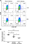
Fig 2. TLR9 is upregulated in non-infected BMDMs.
(A) BMDMs were infected with MOI:0.01 H1N1 and cells were stained for expression of F480, TLR9 and NP by flow cytometry after 24 or 48 h. Flow plots are representative of n = 4–5 samples with similar results in 2 experiments. (B) BMDMs were treated with the TLR9 agonist, ODN 2395, or with the TLR7 agonist, imiquimod (R837), for 24 hours at a concentration of 1μM or 1μg/ml, respectively. RNA was isolated and TLR9 transcript expression was measured by RTqPCR; n = 4; Results analyzed by ANOVA with Tukey post-test.***P<0.001 and are indicative of two similar experiments.
Fig 3
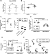
Fig 3. Balb/c and TLR9-/- mice show similar susceptibility to H1N1 and MRSA alone.
(A) Balb/c and TLR9-/- mice were infected with 100 PFU H1N1 and lungs were collected for determination of viral load by plaque assay or (B) by detection of viral M1 gene levels; n = 4-5/group. Panel A was repeated 2 times and panel B is a single experiment done to confirm results of plaque assay. Student’s T-test shows results are not significantly different between genotypes. (C) Total number of cells (left) and percentage of leukocytes (right) in the alveolar compartment of BALB/c and TLR9-/- mice infected with MRSA alone and quantified using a hemocytometer and differential staining; samples were taken by bronchoalveolar lavage 24 hours post-MRSA (7x10⁷ CFUs) infection. (D) Lung bacterial burden and (E) albumin measurements in the BALF 24 and 48 hours post-MRSA (7x10⁷ CFUs) infection in BALB/c and TLR9-/- mice. (F) Cytokine levels in the BALF of BALB/c and TLR9-/- mice infected with MRSA (7x10⁷ CFUs) for 24 hours. Cytokines were measured by ELISA. (C) & (F) Statistics are student T test; ns = non-significant, *P<0.05, **P<0.01, ***P<0.001. Panel C was repeated twice and panel F is a single experiment with n = 4–5 mice/group. (D) and (E) statistics were obtained by one-way analysis of variance with Tukey’s posttest. ****P<0.0001. The 24 h time point was repeated at least 2 times and the 48h time point was a single experiment.
Fig 4
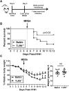
Fig 4. TLR9-/- mice are resistant to secondary bacterial infection.
(A) Sketch of infection model where Balb/c and TLR9-/- mice are infected with 100 PFUs of H1N1 5 days prior to infection with 7x10⁷ CFUs of MRSA (US300). Mice are monitored daily to check for survival. (B) Survival assay of Balb/c (n = 13) and TLR9-/- (n = 13) mice following coinfection. (C) Weight changes (left) and initial weight (right) of all mice used in survival assay. Data are combined from 2 independent experiments with mice matched for weight and sex at start. Statistics for weight changes on each day and initial weight were student T test, non-significant (ns); P<0.05 only on days 8 and 11. Statistics in survival assay were done with the Log-rank (Mantel-Cox), P = 0.03.
Fig 5
Fig 5. TLR9-/- mice experience improved bacterial clearance post-IAV and MRSA coinfection.
(A) Bacterial burden in lungs and (B) albumin levels in BALF from BALB/c and TLR9-/- mice that were infected with IAV (100 PFUs, H1N1) or treated with placebo, PBS, 5 days prior to MRSA (7x10⁷ CFUs) infection; samples were harvested 24 hours post-MRSA infection. (C) Quantification of influenza titers in whole lung of BALB/c and TLR9-/- mice infected with influenza (100 PFUs, H1N1) 5 days prior to MRSA (7x10⁷ CFUs) infection or PBS treatment. (A and C) Statistics were obtained by one-way analysis of variance with Bonferroni’s posttest, Panel B used Tukey’s post-test; ns = non-significant, *P<0.05, **P<0.01, ***P<0.001, ****P<0.0001. All experiments were repeated at least 2 times with similar results.
Fig 6
Fig 6. Higher bacterial clearance in TLR9-/- mice post-IAV and MRSA coinfection is independent of exacerbated levels of IFN-γ.
(A) Cytokine measurement in BALF from BALB/c and TLR9-/- mice that were infected with IAV (100 PFUs, H1N1) 5 days prior to MRSA (7x10⁷ CFUs) infection. (B) Absolute number of lung immune cells post-lung collagenase digestion in BALB/c and TLR9-/- mice that were infected with IAV (100 PFUs, H1N1) 5 days prior to MRSA (7x10⁷ CFUs) infection. Cells were quantified by flow cytometry; gating was as follow: B cells (CD45+CD90.2-CD19+); CD4+ T cells (CD45+CD90.2+CD4+); TH1 (CD45+CD90.2+CD4+,IFN-γ+); CD8+ T cells (CD45+CD90.2+CD4-,CD8+,IFN-γ+); NK cells (CD45+CD90.2+NKP46+,IFN-γ+). (C) Lung bacterial burden and (D) IFN-γ levels in BALB/c and TLR9-/- mice that were treated with 200μg of IFN-γ neutralizing antibody or isotype control and infected with IAV (100 PFUs, H1N1) 5 days prior to MRSA (7x10⁷ CFUs) infection. (A-B) Statistics are student T test between comparative groups; ns = non-significant, *P<0.05, **P<0.01, ***P<0.001, ****P<0.0001. Panels A and B represent single experiments on 4–5 mice/group. (C-D) Statistics were obtained by one-way analysis of variance with Tukey’s posttest; ns = non-significant, *P<0.05, **P<0.01. Panels C and D represent a single experiment due to limited availability of neutralizing antibody.
Fig 7
Fig 7. TLR9-/- lung macrophages have increased phagocytosis, bacterial killing, and iNOS expression post-IAV infection.
(A) Cytokine measurement in BALF from BALB/c and TLR9-/- mice that were infected with IAV (100 PFUs, H1N1) for 5 days n = 5/group (some values are overlapping in the dot plots) for all but IL-10 which was from an experiment with n = 3 TLR9-/- mice and 5 Balb/c mice. (B) Ex vivo MRSA phagocytosis by macrophages isolated by collagenase digestion and adherence purification from mock or H1N1 infected mice on day 5, n = 3. (C) SRA expression analyzed by real-time RT-PCR in monocyte/macrophages isolated on day 5 from H1N1-infected mice, n = 3. (D) Ex vivo MRSA killing assay using adherence selected lung macrophages from BALB/c and TLR9-/- mice that were infected with IAV (100 PFUs H1N1) for 5 days or treated with PBS; n = 8/group in mock infections and n = 3/group in H1N1 infected mice. (E) Quantitative reverse transcriptase-PCR measurement of relative gene expression of iNOS from adherence selected lung macrophages from BALB/c and TLR9-/- mice that were infected with IAV for 5 days or treated with PBS; RNA samples were normalized to their β-actin levels and setting mock-infected Balb/c to 1; n = 3-5/group. (F) BMDMs from Balb/c or TLR9-/- mice were mock-infected for 24 h or were infected with MOI:0.01 H1N1 for 24 or 48 h before RNA was prepared and analyzed for expression of iNOS normalized to their β-actin levels and setting mock-infected Balb/c to 1; n = 3/group. Panels A and C were analyzed by student’s t-test. Panels B, D, E and F were analyzed by two-way ANOVA with Sidak’s multiple comparison test.; Non-significant (ns), *P<0.05, ***P<0.001, ****P<0.0001. Panels A-D are single experiments using multiple mice, panels E and F were repeated two times.
Fig 8
Fig 8. Improved clearance of MRSA post-H1N1 requires loss of TLR9 on non-hematopoietic cells; TLR9 is upregulated on lung epithelial cells post-infection.
(A) Chimeric mice were generated by transplanting bone marrow from Balb/c or TLR9-/- mice into lethally irradiated Balb/c recipients. Following 5 weeks of engraftment, chimeric mice were infected with 7 x 10⁷ CFU MRSA alone or were infected with H1N1 (100 PFU) prior to infection with 7 x 10⁷ MRSA on day 5. Mice were harvested for CFU counts in lungs 24h post-MRSA infection. N = 5 mice/group, single experiment analyzed by ANOVA with Bonferroni post-test. **P<0.01. (B) Adherence purified monocytes and macrophages isolated on day 5 from H1N1-infected mice were treated with control ODN or the TLR9 antagonist ODN2088 at 10 μM concentration for 24h prior to infection with FITC-labeled MRSA for 2 h to measure phagocytosis. Single experiment with N = 10 replicates using cells combined from 3 mice. Data were analyzed by ANOVA with Tukey post-test. **P<0.01, ****P<0.0001. (C) Primary lung epithelial cells were isolated and infected ex vivo with MOI = 0.01 H1N1 or were mock infected for 24 h. Cells were then collected and analyzed for mRNA expression of TLR9. Symbols represent individual wells each with unique mock or H1N1 infection of cells pooled from 3 mice. Data were analyzed by ANOVA with Tukey post-test. *P<0.05, ***P<0.001.

Comment in

References

    1. Iuliano AD, Roguski KM, Chang HH, Muscatello DJ, Palekar R, Tempia S, et al. Estimates of global seasonal influenza-associated respiratory mortality: a modelling study. Lancet (London, England). 2018;391(10127):1285–300. Epub 2017/12/19. 10.1016/s0140-6736(17)33293-2 - DOI - PMC - PubMed
    1. Du Y, Xin L, Shi Y, Zhang TH, Wu NC, Dai L, et al. Genome-wide identification of interferon-sensitive mutations enables influenza vaccine design. Science (New York, NY). 2018;359(6373):290–6. Epub 2018/01/20. 10.1126/science.aan8806 . - DOI - PubMed
    1. McCullers JA. The co-pathogenesis of influenza viruses with bacteria in the lung. Nature reviews Microbiology. 2014;12(4):252–62. Epub 2014/03/05. 10.1038/nrmicro3231 . - DOI - PubMed
    1. Morens DM, Taubenberger JK, Fauci AS. Predominant role of bacterial pneumonia as a cause of death in pandemic influenza: implications for pandemic influenza preparedness. The Journal of infectious diseases. 2008;198(7):962–70. Epub 2008/08/20. 10.1086/591708 - DOI - PMC - PubMed
    1. Dunning J, Blankley S, Hoang LT, Cox M, Graham CM, James PL, et al. Progression of whole-blood transcriptional signatures from interferon-induced to neutrophil-associated patterns in severe influenza. Nature immunology. 2018;19(6):625–35. Epub 2018/05/20. 10.1038/s41590-018-0111-5 - DOI - PMC - PubMed

Publication types

MeSH terms