. 2019 Mar;60(3):406-418.
doi: 10.1111/epi.14653. Epub 2019 Jan 25.

Spectrum of neurodevelopmental disease associated with the GNAO1 guanosine triphosphate-binding region

Collaborators, Affiliations

Spectrum of neurodevelopmental disease associated with the GNAO1 guanosine triphosphate-binding region

McKenna Kelly et al. Epilepsia. 2019 Mar.

Abstract

Objective: To characterize the phenotypic spectrum associated with GNAO1 variants and establish genotype-protein structure-phenotype relationships.

Methods: We evaluated the phenotypes of 14 patients with GNAO1 variants, analyzed their variants for potential pathogenicity, and mapped them, along with those in the literature, on a three-dimensional structural protein model.

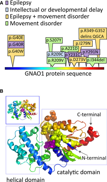
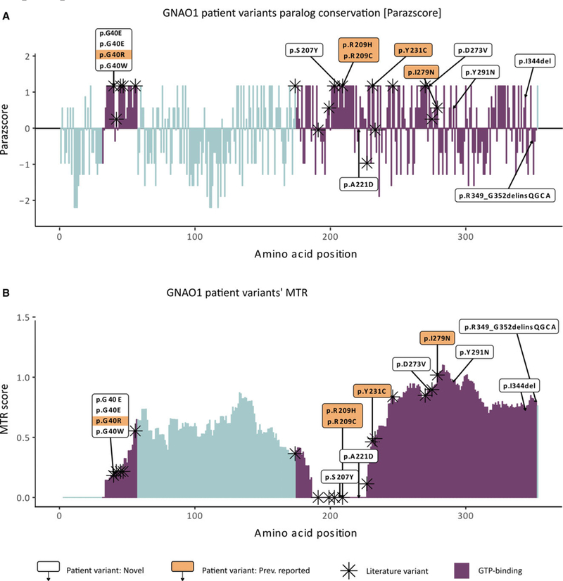
Results: The 14 patients in our cohort, including one sibling pair, had 13 distinct, heterozygous GNAO1 variants classified as pathogenic or likely pathogenic. We attributed the same variant in two siblings to parental mosaicism. Patients initially presented with seizures beginning in the first 3 months of life (8/14), developmental delay (4/14), hypotonia (1/14), or movement disorder (1/14). All patients had hypotonia and developmental delay ranging from mild to severe. Nine had epilepsy, and nine had movement disorders, including dystonia, ataxia, chorea, and dyskinesia. The 13 GNAO1 variants in our patients are predicted to result in amino acid substitutions or deletions in the GNAO1 guanosine triphosphate (GTP)-binding region, analogous to those in previous publications. Patients with variants affecting amino acids 207-221 had only movement disorder and hypotonia. Patients with variants affecting the C-terminal region had the mildest phenotypes.

Significance: GNAO1 encephalopathy most frequently presents with seizures beginning in the first 3 months of life. Concurrent movement disorders are also a prominent feature in the spectrum of GNAO1 encephalopathy. All variants affected the GTP-binding domain of GNAO1, highlighting the importance of this region for G-protein signaling and neurodevelopment.

Keywords: GNAO1; developmental and epileptic encephalopathy; mosaicism; movement disorders.


Conflict of interest statement

DISCLOSURE

The authors have no conflicts of interest to report. We confirm that we have read the Journal’s position on issues involved in ethical publication and affirm that this report is consistent with those guidelines.

Figures

FIGURE 1
FIGURE 1
Patient variants in GNAO1 affect the guanosine triphosphate–binding region. Disease‐associated amino acid residues in the GNAO1 protein localize to the catalytic domain. A, The protein is shown in linear representation, colored using a “rainbow” scheme, starting with blue at the N‐terminal, and ending with red at the C‐terminal. The amino acid substitutions altered by the GNAO1 variants in our series are depicted here, with colors depicting the neurological features present in each case. B, The positions of disease‐associated variants, from this series and the literature, are shown as spheres on the model of the GNAO1 structure. The GNAO1 substrate is also shown as spheres, colored magenta. The same coloring scheme is used in A and B. Patient variants cluster in the region of the catalytic domain (right rectangle), which starts at the very N‐terminal aspect of the sequence, weaves into the helical domain, and returns back to complete the catalytic domain structure. The long helix on the N‐terminal is not shown, because its position in the active conformation of Gα is uncertain, and it carries no reported disease‐associated amino acid positions. Inset (upper left-hand box): The distribution of missense variant amino acids in ExAC covers both structural domains of the protein and does not overlap with the disease‐associated amino acid changes in our series
FIGURE 2
FIGURE 2
GNAO1 patient variant evolutionary conservation and population constrained assessment. A, GNAO1 patient variants’ paralog conservation score (Parazscore) across the linear protein sequence. Comparing the amino acid sequence of the GNAO1 protein to that of paralogous proteins in its gene family, the gene family‐wise paralog conservation is shown for each amino acid of GNAO1 protein sequence. Parazscore values range from negative values, representing less conservation at a given amino acid position, to positive values, representing high conservation, with the highest value indicating that identical amino acids are present in all related proteins. B, GNAO1 patient variants’ missense tolerance ratio (MTR). The score visualizes the tolerance to missense across the GNAO1 protein sequence based on depletion of variants in population controls from the gnomAD database (ExAC v2). MTR values range from 0 (extremely intolerant to missense variant amino acids) to 1.5 (for positions tolerant to missense variant amino acids). For both graphs, patient variant‐related amino acid substitutions not previously reported in the literature (white boxes, n = 9) and patient variant‐related amino acid substitutions previously reported in the literature (shaded boxes, n = 5) are labeled alongside amino acids altered by GNAO1 variants in the literature (asterisks, n = 40). Altogether, 23 amino acid positions are affected. Amino acids belonging to the guanosine triphosphate (GTP)‐binding domain are marked in blue and represent the vast majority of amino acids affected in patients. Intronic variant c.723+1G>A was not included. All patient variants fall within the boundaries of the GTP‐binding domain (blue bars)

References

    1. Scheffer IE, Berkovic S, Capovilla G, et al. ILAE classification of the epilepsies: position paper of the ILAE commission for classification and terminology. Epilepsia 2017;58:512–21. - PMC - PubMed
    1. Nakamura K, Kodera H, Akita T, et al. De novo mutations in GNAO1, encoding a Gαo subunit of heterotrimeric G proteins, cause epileptic encephalopathy. Am J Hum Genet 2013;93:496–505. - PMC - PubMed
    1. EuroEPINOMICS-RES Consortium, Epilepsy Phenome/Genome Project, Epi4K Consortium. De novo mutations in synaptic transmission genes including DNM1 cause epileptic encephalopathies. Am J Hum Genet 2014;95:360–70. - PMC - PubMed
    1. Law CY, Chang ST, Cho SY, et al. Clinical whole‐exome equencing reveals a novel missense pathogenic variant of GNAO1 in a patient with infantile‐onset epilepsy. Clin Chim Acta 2015;451:292–6. - PubMed
    1. Talvik I, Moller RS, Vaher M, et al. Clinical phenotype of de novo GNAO1 mutation: case report and review of literature. Child Neurol Open 2015;2:2329048X15583717. - PMC - PubMed

Publication types

Substances