. 2019 Jan 24;11(2):139.
doi: 10.3390/cancers11020139.

Therapeutic Potential of a Novel αvβ₃ Antagonist to Hamper the Aggressiveness of Mesenchymal Triple Negative Breast Cancer Sub-Type

Affiliations

Therapeutic Potential of a Novel αvβ₃ Antagonist to Hamper the Aggressiveness of Mesenchymal Triple Negative Breast Cancer Sub-Type

Billy Samuel Hill et al. Cancers (Basel). .

Abstract

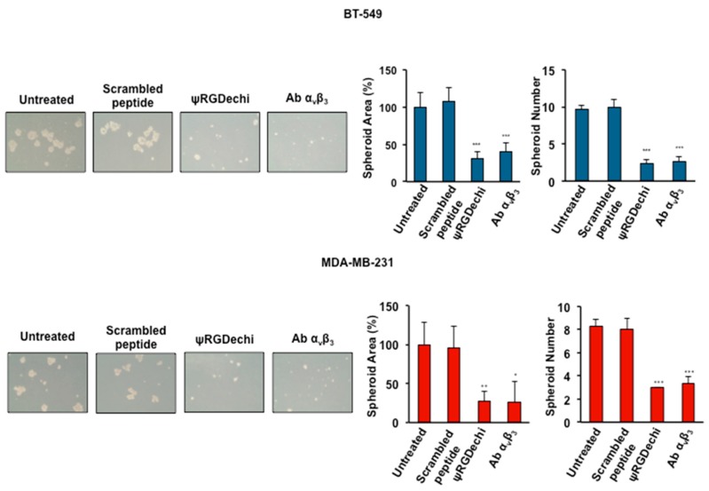
The mesenchymal sub-type of triple negative breast cancer (MES-TNBC) has a highly aggressive behavior and worse prognosis, due to its invasive and stem-like features, that correlate with metastatic dissemination and resistance to therapies. Furthermore, MES-TNBC is characterized by the expression of molecular markers related to the epithelial-to-mesenchymal transition (EMT) program and cancer stem cells (CSCs). The altered expression of αvβ₃ integrin has been well established as a driver of cancer progression, stemness, and metastasis. Here, we showed that the high levels of αvβ₃ are associated with MES-TNBC and therefore exploited the possibility to target this integrin to reduce the aggressiveness of this carcinoma. To this aim, MES-TNBC cells were treated with a novel peptide, named ψRGDechi, that we recently developed and characterized for its ability to selectively bind and inhibit αvβ₃ integrin. Notably, ψRGDechi was able to hamper adhesion, migration, and invasion of MES-TNBC cells, as well as the capability of these cells to form vascular-like structures and mammospheres. In addition, this peptide reversed EMT program inhibits mesenchymal markers. These findings show that targeting αvβ₃ integrin by ψRGDechi, it is possible to inhibit some of the malignant properties of MES-TNBC phenotype.

Keywords: cell migration and invasion; epithelial-mesenchymal transition; stemness; triple-negative breast cancer; αvβ3 integrin; ψRGDechi.


Conflict of interest statement

The authors declare no conflict of interest.

Figures

Figure 1
Figure 1
αvβ3 integrin expression is associated with triple-negative breast cancer (TNBC) mesenchymal subtype. (A) Analysis of αV (ITGAV) and β3 (ITGB3) mRNA expression and their correlation to clinical, molecular, and cell phenotype, was performed on 198 TNBC tumors from MD Anderson Cancer Center (dataset: GSE76124). All correlations were assessed by Pearson’s χ2 test or with one-way analysis of variance (ANOVA), through the R2 platform (Academic Medical Center, Netherlands) and presented in BoxPlots and subdivided into TNBC sub-types. BLIA: Basal-like immune-activated; BLIS: Basal-like immune-suppressed; LAR: Luminal-androgen receptor; MES: mesenchymal. *** p < 0.0001. (B) MES-TNBC cells (1 × 10⁶) were incubated with FITC mouse antibody against human integrin αvβ3 (LM609) and analyzed using a FACSCalibur System (BD Biosciences, San Jose, CA, USA). Isotype-matched antibodies were used as controls.
Figure 2
Figure 2
ψRGDechi inhibits MES-TNBC cell adhesion. BT-549 and MDA-MB-231 cells (8 × 10⁴ cells/well) were suspended and mixed in a binding solution with ψRGDechi (from 0.1 to 50 µM) or anti-αvβ3 antibody LM609 (10 μg/mL) (Millipore, Burlington, MA, USA), for 30 min at room temperature, then seeded on plates pre-coated with 5 μg/mL vitronectin and allowed to attach for 2 h. The non-adherent cells were removed using PBS, and the attached cells were stained using a 0.1% crystal violet solution in 25% methanol for 30 minutes. All the results are expressed as the percentage of adherent cells considering the untreated as 100%. Bars depict mean ± SD of three independent experiments. *** p < 0.0001; ** p < 0.001.
Figure 3
Figure 3
ψRGDechi inhibits MES-TNBC cell migration and cell wound healing ability. (A) Cell migration was performed using a 24-well Boyden chamber. BT-549 and MDA-MB-231 cells (0.5 × 10⁵ cells/well) were re-suspended in 100 µL of serum-free medium in presence or absence of different concentration of ψRGDechi (50 µM, 10 µM, 1 µM), scrambled-peptide (50 µM), and blocking anti-αvβ3 antibody LM609 (10 μg/mL) and seeded in the upper chamber. Medium containing 1% FBS or 10% FBS was added to the lower chamber as a chemoattractant. The results are expressed as the percentage of migrating cells considering the untreated control sample (1% FBS) as 100%. B) Both cell lines were grown as confluent monolayers in 6-well plates were scratched with pipette tips to create wounds. After removal of detached cells, medium containing 1% FBS, 10% FBS, ψRGDechi (10 µM), scrambled-peptide (10 µM) and blocking anti-αvβ3 antibody LM609 (10 μg/mL), were added to cells. Each scratch area was photographed at 0, 24, and 48 h. The distance between the edges of the scratch was measured by ImageJ, the average distance was quantified and the extent of wound closure was determined as follows: wound closure (%) = 1 − (wound width tx/wound width t0) × 100. Bars depict mean ± SD of three independent experiments. *** p < 0.0001; ** p < 0.001; * p < 0.01.
Figure 4
Figure 4
ψRGDechi inhibits MES-TNBC cell ability to invade extracellular matrix. The invasion assay was performed using the Boyden chamber with membranes (8 µm pores) coated with Matrigel. BT-549 and MDA-MB-231 (1 × 10⁵) cells were harvested, suspended in serum free medium alone or containing, ψRGDechi (10 µM), scrambled peptide (10 µM) and anti-αvβ3 antibody (10 µg/mL) and placed in the top chamber. In the lower chamber medium containing 1% FBS or 10% FBS was added and used as a chemoattractant. All experiments were performed at least three times and the results are expressed as the percentage of invasive cells considering the untreated control sample (1% FBS) as 100%. Bars depict mean ± SD of three independent experiments. *** p < 0.0001.
Figure 5
Figure 5
ψRGDechi inhibits MES-TNBC cell ability to form vascular-like structures. Harvested MDA-MB-231 (1 × 10⁵) and BT-549 (8 × 10⁴) cells were suspended in 100 µL medium containing 2% FBS in presence or absence of ψRGDechi (10 µM), scrambled peptide (10 µM) and anti-αvβ3 antibody (10 µg/mL). Treated cells were then seeded into 24-well plates pre-coated with 80 µL/well Matrigel and incubated at 37 °C and 5% CO2 for 24 h. Representative images were taken at 10× and average of the number of complete loops was calculated from 3–5 random fields by a macro made with ImageJ software. Bars depict mean ± SD of three independent experiments. ** p < 0.001.
Figure 6
Figure 6
ψRGDechi inhibits MES-TNBC cell ability to form spheroids. BT-549 and MDA-MB-231 cells (5 × 10⁴/well) were seeded in Ultra-Low attachment 6-multiwell-plates and grown in serum-free DMEM supplemented with B27, bFGF (20 ng/mL) EGF (10 ng/mL). Cells were incubated at 37 °C with 5% CO2 for 7 days. Spheroid formation was analyzed under a phase-contrast microscopy and size and number of formed spheroids was calculated using imageJ. Bars depict mean ± SD of three independent experiments. *** p < 0.0001; ** p < 0.001; * p < 0.01.
Figure 7
Figure 7
ψRGDechi inhibits EMT program in MES-TNBC cells. Analysis by western blot of Vimentin, N-cadherin, Slug, E-cadherin, p-AKT and AKT levels in untreated, scramble-peptide treated and ψRGDechi treated BT-549 and MDA-MB-231 cells. Actin and Tubulin were used as loading control. Values below the blot indicate signal levels relative to the untreated cells, which were arbitrarily set to 1. Representative data from one of three experiments are shown.
Figure 8
Figure 8
Schematic model illustrating that the blocking of αvβ3 integrin by ψRGDechi reverses the EMT program and reduces migration, invasion, vascular mimicry and stemness in MES-TNBC.

References

    1. Bianchini G., Balko J.M., Mayer I.A., Sanders M.E., Gianni L. Triple-negative breast cancer: Challenges and opportunities of a heterogeneous disease. Nat. Rev. 2016;13:674–690. doi: 10.1038/nrclinonc.2016.66. - DOI - PMC - PubMed
    1. Lehmann B.D., Bauer J.A., Chen X., Sanders M.E., Chakravarthy A.B., Shyr Y., Pietenpol J.A. Identification of human triple-negative breast cancer subtypes and preclinical models for selection of targeted therapies. J. Clin. Investig. 2011;121:2750–2767. doi: 10.1172/JCI45014. - DOI - PMC - PubMed
    1. Lehmann B.D., Jovanović B., Chen X., Estrada M.V., Johnson K.N., Shyr Y., Moses H.L., Sanders M.E., Pietenpol J.A. Refinement of Triple-Negative Breast Cancer Molecular Subtypes: Implications for Neoadjuvant Chemotherapy Selection. PLoS ONE. 2016;11:e0157368. doi: 10.1371/journal.pone.0157368. - DOI - PMC - PubMed
    1. Burstein M.D., Tsimelzon A., Poage G.M., Covington K.R., Contreras A., Fuqua S.A., Savage M.I., Osborne C.K., Hilsenbeck S.G., Chang J.C., et al. Comprehensive genomic analysis identifies novel subtypes and targets of triple-negative breast cancer. Clin. Cancer Res. 2015;21:1688–1698. doi: 10.1158/1078-0432.CCR-14-0432. - DOI - PMC - PubMed
    1. Yu K.D., Zhu R., Zhan M., Rodriguez A.A., Yang W., Wong S., Makris A., Lehmann B.D., Chen X., Mayer I., et al. Identification of prognosis-relevant subgroups in patients with chemoresistant triple-negative breast cancer. Clin. Cancer Res. 2013;19:2723–2733. doi: 10.1158/1078-0432.CCR-12-2986. - DOI - PMC - PubMed

LinkOut - more resources