. 2019 Jan 25;19(1):23.
doi: 10.1186/s12866-019-1392-z.

Interaction of different Chlamydiae species with bovine spermatozoa

Affiliations

Interaction of different Chlamydiae species with bovine spermatozoa

Thomas Eckert et al. BMC Microbiol. .

Abstract

Background: Interaction of spermatozoa and Chlamydiae spp. might contribute to reduced fertility in cattle. To proof this hypothesis, bovine semen was incubated with viable or heat inactivated Chlamydia (C.) abortus or psittaci (Multiplicity of infection = 1) and sperm motility was monitored with a computer-assisted sperm analyzer over 24 h. Additionally, the interaction with the spermatozoa was further investigated by means of light and transmission electron microscopy (TEM).

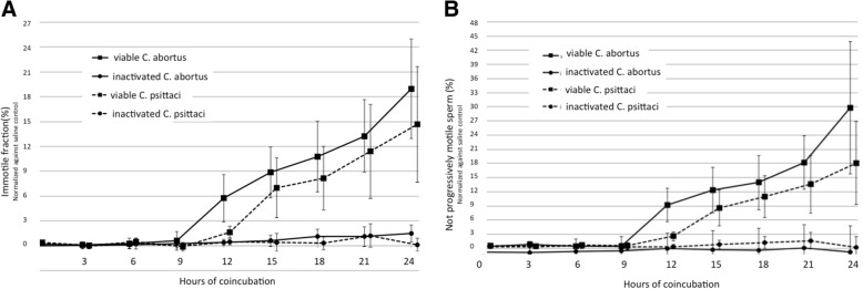
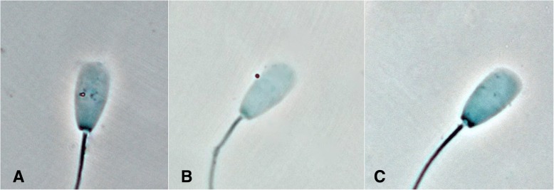
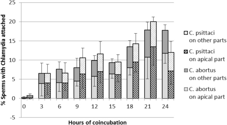
Results: Only viable Chlamydiae of both species decreased sperm motility and this only after about 9 h. Taking binding rates into account, the loss of sperm motility after about 9 h could likely be a consequence of Chlamydiae attachment to the spermatozoa. About two thirds of the Chlamydiae elementary bodies were bound to the front third of the sperm, the acrosomal region. No inclusions of Chlamydiae in spermatozoa were observed in TEM after 2 h co-incubation.

Conclusions: As initial motility was not affected following co-incubation of viable Chlamydiae and bovine sperm, it seems likely that sperm could serve as a carrier/vehicle for Chlamydiae facilitating cervical passage of Chlamydiae spp. in cattle. Additionally, our results suggest that spermatozoa carrying Chlamydiae may have no initial disadvantage in reaching the oviduct, but are immotile at the time of ovulation what might have an impact on fertilization capacities of the individual sperm. Consequently, high concentrations of the investigated Chlamydiae in the seminal plasma or female genital tract might play a role in reduced fertility in cattle.

Keywords: CASA (computer assisted sperm analysis); Cattle; Chlamydiae; Semen motility.


Conflict of interest statement

Ethics approval

This study was carried out in strict accordance with the recommendations of the Guide for the Care and Use of Laboratory Animals of the National Institutes of Health. The housing facilities and the protocol were approved by the Regierungspräsidium Giessen, Germany (AZ No. V 54-19c2015h02 GI 18/14 Nr. A 27/2012).

Consent for publication

Not applicable.

Competing interests

The authors declare no conflict of interest. I confirm that I have read BioMed Central’s guidance on competing interests and have included a statement indicating that none of the authors have any competing interests in the manuscript.

Publisher’s Note

Springer Nature remains neutral with regard to jurisdictional claims in published maps and institutional affiliations.

Figures

Fig. 1
Fig. 1
Viable Chlamydia abortus und psittaci signicantly decrease a. total and b. progressive motility of bovine spermatozoa. Percentage of immotile bovine sperm (normalized against saline control) coincubated at 35 °C with viable and heat-inactivated Chlamydia (MOI = 1) for 24 h. Results are depicted as mean values ± SD of 5 replicates. Motility of spermatozoa co-incubated with viable Chlamydiae spp. was significantly reduced from 9 h onwards compared to motility of spermatozoa incubated with heat inactivated Chlamydiae (p < 0.0001)
Fig. 2
Fig. 2
Light microscopical evaluation clearly confirms binding of C. abortus and psittaci to bovine spermatozoa. The orange particles represent chlamydial particles (a. C. abortus; b. C. psittaci) bound to a bovine spermatozoon. The particles look orange due to the use of a blue filter. In the negative control (c.), no similar particles are visible (magnification: × 1000)
Fig. 3
Fig. 3
C. abortus and psittaci can attach to bovine spermatozoa. The apical area of the sperm head seems to be the preferred location for attachment. Attachment of Chlamydia spp. (C. abortus, C. psittaci,) to the apical (acrosomal) area and other parts of bovine spermatozoa during 24 h of coincubation (MOI = 1) at 35 °C. Results are depicted as mean values of 4 replicates of 50 sperms ± SD. [The top error bar indicates the error bar of “the other parts” for the respective Chlamydia spp. (only positive SD presented); the lower error bar indicates the error bar of “the apical part” (only negative SD presented).] Approximately 60% of Chlamydiae spp. particles are attached to the apical area corresponding to the acrosomal region
Fig. 4
Fig. 4
Transmission electron microscopic images showing Chlamydia-sperm interaction Exemplary chlamydial particles are marked. a. Chlamydial particles in Buffalo-Green-Monkey-Kidney cell culture after harvesting, size indicating elementary bodies, additionally cell debris visible and b. a Chlamydia spp. particle, resembling an elementary body localised at the apical part of the sperm head (all magnifications M12000, bar indicates 0.6 μm)

Similar articles

Cited by

References

    1. Eggert-Kruse W, Gerhard I, Naher H, Tilgen W, Runnebaum B. Chlamydial infection-a female and/or male infertility factor? Fertil Steril. 1990;53:1037–1043. doi: 10.1016/S0015-0282(16)53581-X. - DOI - PubMed
    1. Wolner-Hanssen P, Mardh PA, Moller B, Westrom L. Endometrial infection in women with chlamydial salpingitis. Sex Transm Dis. 1982;9:84–88. doi: 10.1097/00007435-198204000-00006. - DOI - PubMed
    1. Wolner-Hanssen P, Westrom L, Mardh PA. Perihepatitis and chlamydial salpingitis. Lancet. 1980;1:901–903. doi: 10.1016/S0140-6736(80)90838-7. - DOI - PubMed
    1. Reinhold P, Sachse K, Kaltenboeck B. Chlamydiaceae in cattle: commensals trigger organisms or pathogens? Vet J. 2011;189:257–267. doi: 10.1016/j.tvjl.2010.09.003. - DOI - PubMed
    1. Schautteet K, Vanrompay D. Chlamydiaceae infections in pig. Vet Res. 2011;42:29. doi: 10.1186/1297-9716-42-29. - DOI - PMC - PubMed

Publication types