. 2019 Jan 25;17(1):9.
doi: 10.1186/s12964-018-0316-0.

Epigenetic inactivation of HOXD10 is associated with human colon cancer via inhibiting the RHOC/AKT/MAPK signaling pathway

Affiliations

Epigenetic inactivation of HOXD10 is associated with human colon cancer via inhibiting the RHOC/AKT/MAPK signaling pathway

Yu-Hong Yuan et al. Cell Commun Signal. .

Abstract

Background: To examine the influence of HOXD10 on the metabolism and growth of colon carcinoma cells by suppressing the RHOC/AKT/MAPK pathway.

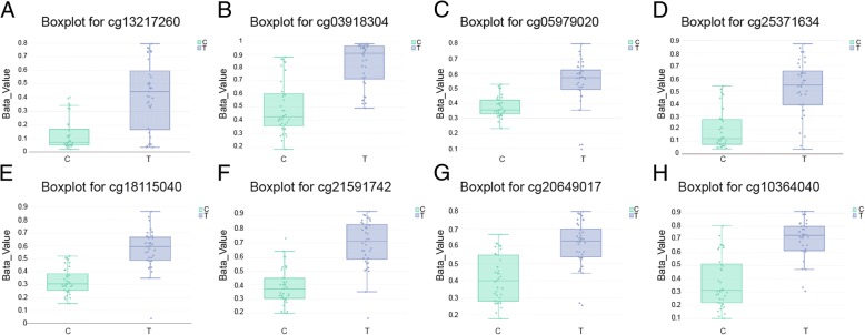
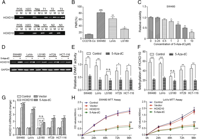
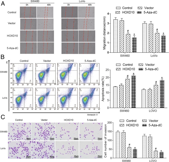
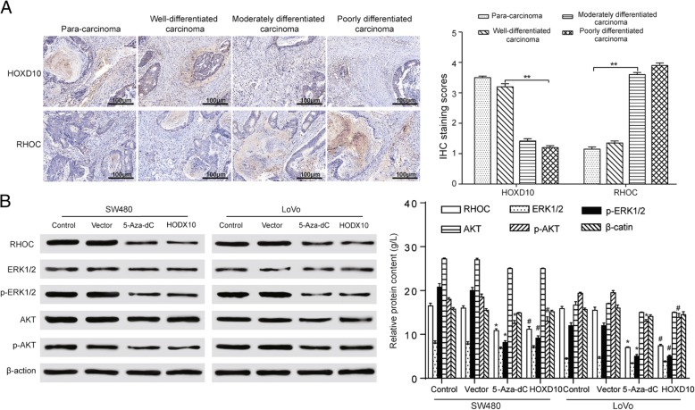
Methods: Thirty-seven paired colon cancer and its adjacent samples from The Cancer Genome Atlas (TCGA) were analyzed. Chip Analysis Methylation Pipeline (ChAMP) analysis was employed for differential methylated points (DMPs) and the differential methylation regions (DMRs) screening. The HOXD10 mRNA expression and DNA methylation levels were detected by RT-PCR. The Cell proliferation, migration, invasion and apoptosis were respectively measured by MTT assay, transwell assay, wound healing assay and flow cytometry assay in carcinoma cell lines after treated with 5-aza-2'-deoxycytidine (5-Aza-dC) or transfected with HOXD10-expressing plasmid. The expression of HOXD10 and RHOC was revealed by immunohistochemistry in disparate differentiation colon carcinoma tissues, and the dephosphorylation of AKT and MAPK pathways were detected by RT-PCR and western blot.

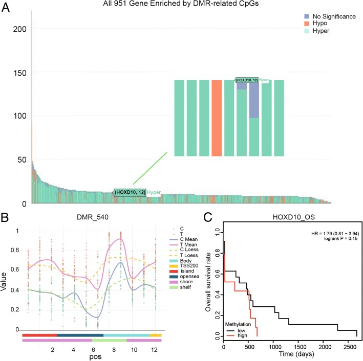
Results: The bioinformatics analysis demonstrated that HOXD10 was hypermethylated and low-expressed in colorectal cancer tissues. The detection of RT-PCR indicated the similar results in colorectal cancer cell lines and tissues. The induction of demethylation was recovered by treatment with 5-Aza-dC and the HOXD10 in colorectal cancer cell lines was re-expressed by transfection with a HOXD10 expression vector. The demethylation or overexpression of HOXD10 suppressed proliferation, migration, invasion and promoted apoptosis in colorectal cancer cells. HXOD10 suppressed the tumor growth and detected an opposite trend of protein RHOC. AKT and MAPK pathways were notably inactivated after the dephosphorylation due to the overexpression of HOXD10.

Conclusions: HOXD10 was suppressed in colon adenocarcinoma cells, which down-regulated RHOC/AKT/MAPK pathway to enhance colon cancer cells apoptosis and constrain the proliferation, migration and invasion.

Keywords: 5-Aza-dC; Colon cancer; HOXD10; Methylation; RHOC/AKT/MAPK pathway.


Conflict of interest statement

Ethics approval and consent to participate

All procedures performed in studies involving human participants were in accordance with the ethical standards of Sun Yat-sen Memorial Hospital. Written informed consents were obtained from all individual participants included in the study.

Consent for publication

The authors consent for publication.

Competing interests

The authors declare that they have no competing interests.

Publisher’s Note

Springer Nature remains neutral with regard to jurisdictional claims in published maps and institutional affiliations.

Figures

Fig. 1
Fig. 1
Distribution of top 1000 differentially methylated imprinted CpG sites. a Distribution of top 1000 differentially methylated imprinted CpG sites according to CpG islands (shores, shelves, islands, and opensea). b Distribution of top 1000 differentially methylated imprinted CpG sites according to the gene position (1stExon, 3’ UTRs or 5’ UTRs, body, IGR, TSS1500 and TSS200). c Distribution of top 1000 differentially methylated imprinted CpG sites according to the information of genetic and epigenetic annotation
Fig. 2
Fig. 2
Genome-wide methylation data was from TCGA for 37 available COAD (Colon Adenocarcinoma) tumor/surrounding pairs. a Heatmap of top 1000 differentially methylated imprinted CpG sites. b Density plot of methylated DNA intensity for each sample. The quality of the data for each specimen was visualized using a density plot, displaying different β-value distributions (0 indicating unmethylated sites, 1 indicating fully methylated sites). c Multi-dimensional scaling (MDS) plot showing differential clustering of control vs. tumor tissues
Fig. 3
Fig. 3
HOXD10 hypermethylation was associated with lower mRNA expression. a Differential mRNA expression analysis using paired tumor/surrounding tissues identified top 40 differentially mRNA genes. Lower HOXD10 mRNA expression in tumor tissues compare to paired normal tissues. b Differential methylation analysis using paired tumor/surrounding tissues identified top 40 differentially methylated genes. Higher HOXD10 methylation in tumor tissues compare to the counterpart
Fig. 4
Fig. 4
HOXD10 was enriched by CpGs by DMR (differentially methylated region) analysis. a The bar chart shows that HOXD10 was hyper-enriched by DMR-related CpGs among all 951 genes. b A differentially methylated region (DMR_540) of HOXD10 demonstrates that the expression of this gene in tumor group apparently higher than normal group. c Kaplan-Meier plot of the overall survival rate of COAD patients based on HOXD10 -high or -low methylation
Fig. 5
Fig. 5
Eight CpG sites for HOXD10 which presented in boxplot displayed an increased methylation in the tumor group. Boxplot for (a) cg 13,217,260, (b) cg 03918304, (c) cg 05979020, (d) cg 25,371,634, (e) cg 18,115,040, (f) cg 21,591,742, (g) cg 20,649,017, (h) cg 10,364,040
Fig. 6
Fig. 6
5-Aza-dC induced demethylation and re-expression of silenced HOXD10. a Higher HOXD10 methylation in tumor tissues compare to paired normal tissues. ‘Pos’ represented the positive controls for the methylated (M) and unmethylated (U) allele. The normal peripheral lymphocytes DNA was used as negative control. b HOXD10 was confirmed to be hypermethylated in colon adenocarcinoma cell lines SW480 and LoVo compared with normal colon cell line CCD-18Co cell line. The change of DNA methylation level was minimal in LS180 (COAD cell line) compared with CCD-18Co cells. * P < 0.05, ** P < 0.01, *** P < 0.001, compared with the CCD-18Co cell lines. c Minimum effective dose of 5-Aza-dC was determined by MTT. 1 μM showed difference. * P < 0.05, ** P < 0.01, compared with the 0 μM group. d The methylation level of HOXD10 decreased after the treatment with demethylation agent 5-Aza-dC in SW480, LoVo, LS180, HT29 and HCT-116 cell lines. e Relative DNMT activity was decreased after treatment of the cells with 5-Aza-dC (1 μM) for 72 h. Compared with the control group, ** P < 0.01. f 5-Aza-dC (1 μM) treatment decreased HOXD10 methylation in SW480, LoVo, LS180, HT29 and HCT-116 cell lines. Compared with the control group, *** P < 0.001. g The mRNA expression of HOXD10 in SW480, LoVo, LS180, HT29 and HCT-116 cell lines was higher after treatment with 5-Aza-dC (1 μM) for 72 h or overexpression of HOXD10. *** indicated P < 0.001 compared with the control group; ## indicated P < 0.01 compared with the vector congtrol group. h Cell proliferation was suppressed in SW480 and LoVo cells after treatment with 5-Aza-dC (1 μM) for 72 h or overexpression of HOXD10, determined by MTT assay. Compared with the control group, *** P < 0.001. Compared with the vector control group, ###P < 0.001
Fig. 7
Fig. 7
HOXD10 re-expression inhibited cell migration, invasion and promoted the cell apoptosis. a HOXD10 reduced the migration rates of SW480 and LoVo cells in scratch wound-healing assay, and photographs were taken at 0, 48 h after the wound was made (left). Statistical plot of the average number of migrated SW480 and LoVo cells in each group (right). * indicated P < 0.05 in comparison with the control group and # indicated P < 0.05 in comparison with the vector control group. b The result of flow cytometry showed HOXD10 overexpression induced SW480 and LoVo cells apoptosis (left). Statistical plot displayed percentages of apoptosis in SW480 and LoVo cells (right), *P < 0.05;#P < 0.05. c Overexpression of HOXD10 significantly decreased the invasive potential of both SW480 and LoVo cell lines through Matrigel invasion Transwell assay (left). Statistical plot of the average number of invaded SW480 and LoVo cells in each group (right). The graph showed the mean ± SD. *P < 0.05; #P < 0.05
Fig. 8
Fig. 8
The expression of HOXD10 displayed contrary tendency with protein RHOC. a HOXD10 stainings were gradually weakening while RHOC expression showed a reverse trend in para-carcinoma (× 100), well- (× 400), moderately- (× 400) and poorly-differentiated carcinoma (× 400), observed by immunohistochemistry (left). Statistical plot illustrates IHC staining scores in SW480 and LoVo cells (right). b Suppressed protein expression levels of RHOC, phosphorylated AKT, and phosphorylated ERK (1/2), and unchanged expression of AKT and ERK (1/2) in SW480 and LoVo cells compared with the control groups (left). Statistical plot showed the relative protein expression in RHOC/AKT/ERK in SW480 and LoVo cells (right). *P < 0.05, **P < 0.01 compared with the control group; #P < 0.05, ##P < 0.01 compared with the vector control group

References

    1. Rafiemanesh H, Mohammadian-Hafshejani A, Ghoncheh M, et al. Incidence and mortality of colorectal Cancer and relationships with the human development index across the world. Asian Pac J Cancer Prev. 2016;17:2465–2473. doi: 10.7314/APJCP.2016.17.3.1393. - DOI - PubMed
    1. Takiyama A, Nozawa H, Ishihara S, et al. Secondary metastasis in the lymph node of the bowel invaded by colon cancer: a report of three cases. World J Surg Oncol. 2016;14:273. doi: 10.1186/s12957-016-1026-y. - DOI - PMC - PubMed
    1. Jung KW, Won YJ, Oh CM, et al. Prediction of Cancer incidence and mortality in Korea, 2016. Cancer Res Treat. 2016;48:451–457. doi: 10.4143/crt.2016.092. - DOI - PMC - PubMed
    1. Chen W, Zheng R, Zeng H, et al. Annual report on status of cancer in China, 2011. Chin J Cancer Res. 2015;27:2–12. doi: 10.1186/s40880-015-0001-2. - DOI - PMC - PubMed
    1. Bishehsari F, Mahdavinia M, Vacca M, et al. Epidemiological transition of colorectal cancer in developing countries: environmental factors, molecular pathways, and opportunities for prevention. World J Gastroenterol. 2014;20:6055–6072. doi: 10.3748/wjg.v20.i20.6055. - DOI - PMC - PubMed

Publication types

MeSH terms