. 2019 Jan 25;17(1):18.
doi: 10.1186/s12951-019-0451-9.

Mitochondria-targeted cyclosporin A delivery system to treat myocardial ischemia reperfusion injury of rats

Affiliations

Mitochondria-targeted cyclosporin A delivery system to treat myocardial ischemia reperfusion injury of rats

Chang-Xiong Zhang et al. J Nanobiotechnology. .

Abstract

Background: Cyclosporin A (CsA) is a promising therapeutic drug for myocardial ischemia reperfusion injury (MI/RI) because of its definite inhibition to the opening of mitochondrial permeability transition pore (mPTP). However, the application of cyclosporin A to treat MI/RI is limited due to its immunosuppressive effect to other normal organ and tissues. SS31 represents a novel mitochondria-targeted peptide which can guide drug to accumulate into mitochondria. In this paper, mitochondria-targeted nanoparticles (CsA@PLGA-PEG-SS31) were prepared to precisely deliver cyclosporin A into mitochondria of ischemic cardiomyocytes to treat MI/RI.

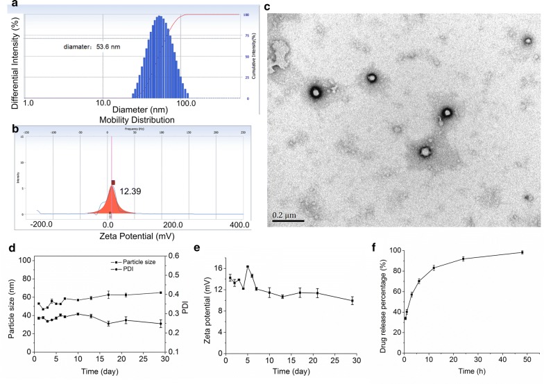
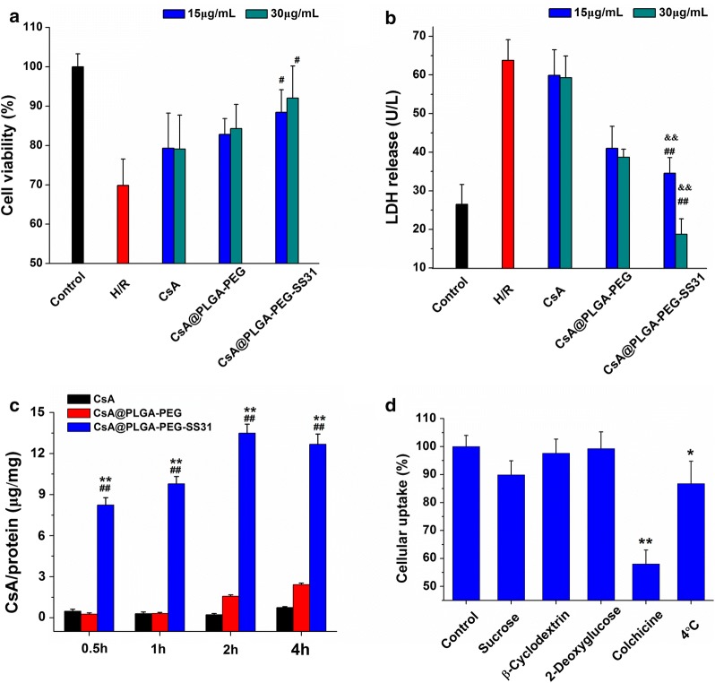
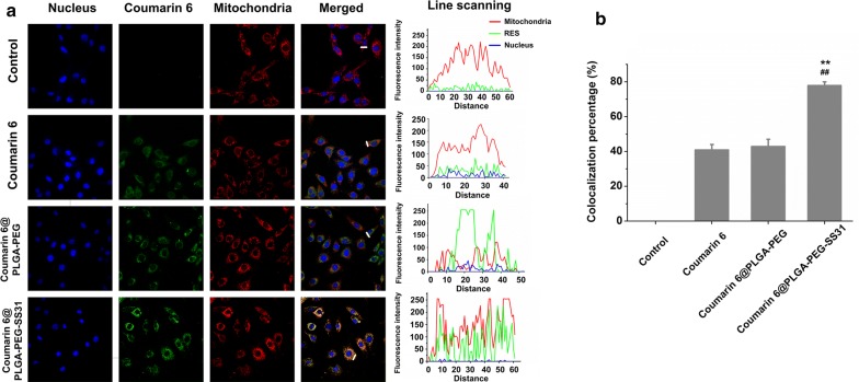
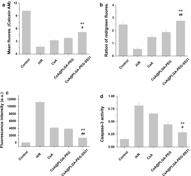
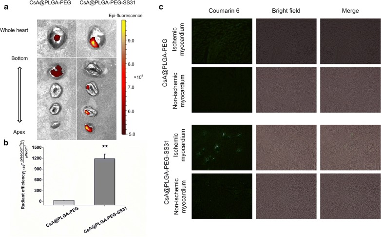
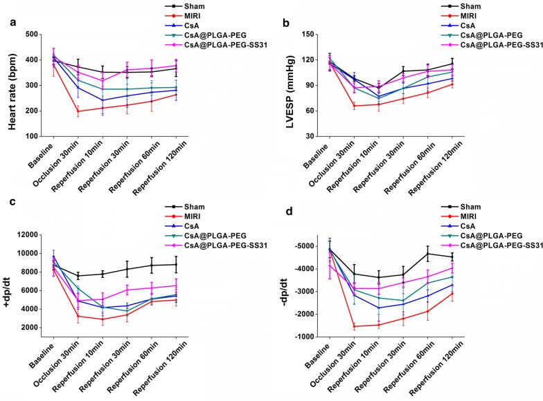
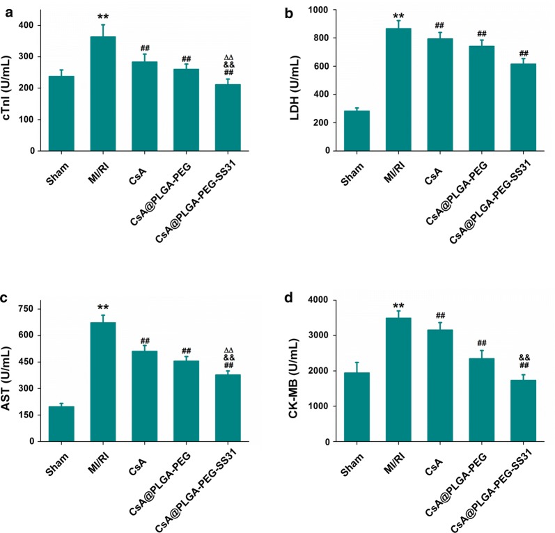
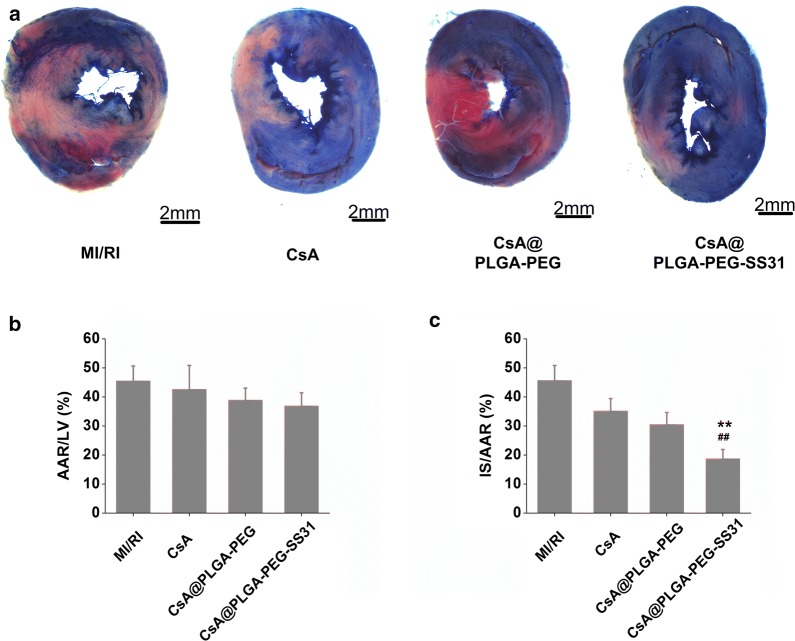
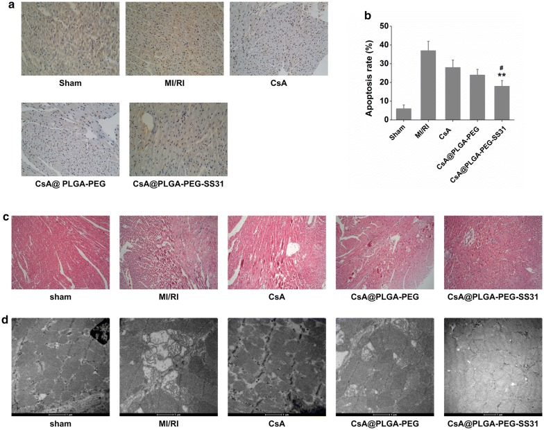
Results: CsA@PLGA-PEG-SS31 was prepared by nanoprecipitation. CsA@PLGA-PEG-SS31 showed small particle size (~ 50 nm) and positive charge due to the modification of SS31 on the surface of nanoparticles. CsA@PLGA-PEG-SS31 was stable for more than 30 days and displayed a biphasic drug release pattern. The in vitro results showed that the intracellular uptake of CsA@PLGA-PEG-SS31 was significantly enhanced in hypoxia reoxygenation (H/R) injured H9c2 cells. CsA@PLGA-PEG-SS31 delivered CsA into mitochondria of H/R injured H9c2 cells and subsequently increased the viability of H/R injured H9c2 cell through inhibiting the opening of mPTP and production of reactive oxygen species. In vivo results showed that CsA@PLGA-PEG-SS31 accumulated in ischemic myocardium of MI/RI rat heart. Apoptosis of cardiomyocyte was alleviated in MI/RI rats treated with CsA@PLGA-PEG-SS31, which resulted in the myocardial salvage and improvement of cardiac function. Besides, CsA@PLGA-PEG-SS31 protected myocardium from damage by reducing the recruitment of inflammatory cells and maintaining the integrity of mitochondrial function in MI/RI rats.

Conclusion: CsA@PLGA-PEG-SS31 exhibited significant cardioprotective effects against MI/RI in rats hearts through protecting mitochondrial integrity, decreasing apoptosis of cardiomyocytes and myocardial infract area. Thus, CsA@PLGA-PEG-SS31 offered a promising therapeutic method for patients with acute myocardial infarction.

Keywords: Cyclosporin A; Mitochondria-targeted peptide; Mitochondrial permeability transition pore; Mitochondrial targeting; Myocardial ischemia/reperfusion injury.


Figures

Fig. 1
Fig. 1
Physicochemical and in vitro characterization of CsA@PLGA-PEG-SS31. a Size distribution of CsA@PLGA-PEG-SS31. b Zeta potential of CsA@PLGA-PEG-SS31. c TEM image of CsA@PLGA-PEG-SS31. d The particle size of CsA@PLGA-PEG-SS31 in deionized water. e The zeta potential of CsA@PLGA-PEG-SS31 in deionized water. f In vitro drug release profiles of CsA@PLGA-PEG-SS31 in 40% ethanol
Fig. 2
Fig. 2
a Cell viability of H/R injury H9c2 cell after treatment with different concentration of CsA, CsA@PLGA-PEG and CsA@PLGA-PEG-SS31. #p < 0.05 versus CsA group at the same concentration. b LDH release from H/R injury H9c2 cell after treatment with different concentration of CsA, CsA@PLGA-PEG and CsA@PLGA-PEG-SS31. ##p < 0.01 versus CsA group at the same concentration, &&p < 0.01 versus CsA@PLGA-PEG group at the same concentration. c Cellular uptake of CsA, CsA@PLGA-PEG and CsA@PLGA-PEG-SS31 in H/R injured H9c2 cells (determined by HPLC). ##p < 0.01 versus CsA group at the same time point, **p < 0.01 versus CsA@PLGA-PEG group at the same time point. d Relative uptake efficiency of CsA@PLGA-PEG-SS31 on H/R injured H9c2 cells at the presence of various endocytosis inhibitors, ATP depletion or 4 °C. **p < 0.01, *p < 0.05 versus control group
Fig. 3
Fig. 3
The intracellular trafficking of CsA@PLGA-PEG-SS31 in H/R injured H9c2 cells. a The mitochondrial distribution and the fluorescence intensity after free coumarin 6, coumarin 6 labeled CsA@PLGA-PEG (coumarin 6@PLGA-PEG), and coumarin 6 labeled CsA@PLGA-PEG-SS31 (coumarin 6@PLGA-PEG-SS31) were incubated with H/R injured H9c2 cells for 4 h (60 × 10). The fluorescence intensity was graphed according to the white line of merged fluorescence images. b Colocalization efficiency of coumarin 6 after free coumarin 6, coumarin 6 labeled CsA@PLGA-PEG (coumarin 6@PLGA-PEG) and coumarin 6 labeled CsA@PLGA-PEG-SS31 (coumarin 6@PLGA-PEG-SS31) incubated with H/R injured H9c2 cells for 4 h (×60). **p < 0.01 versus coumarin 6 group, ##p < 0.01 versus coumarin 6@PLGA-PEG group
Fig. 4
Fig. 4
The protective effects of CsA@PLGA-PEG-SS31 on H/R injured H9c2 cells. a Calcien AM fluorescence intensity measured in H/R injured H9c2 cells by flow cytometry. b The ratio of red/green fluorescence intensity measured by fluoro-spectrophotometer in H/R injured H9c2 cells after JC-1staining. c MitoSOX fluorescence intensity measured in H/R injured H9c2 cells by flow cytometry. d Caspase 3 activity measured by fluoro-spectrophotometer in H/R injured H9c2 cells. **p < 0.01 versus CsA group; #p < 0.05, ##p < 0.01 versus CsA@PLGA-PEG group
Fig. 5
Fig. 5
Representative ex vivo fluorescence images (a) and quantitative analysis (b) of cy7.5 labelled CsA@PLGA-PEG and cy7.5 labelled CsA@PLGA-PEG-SS31 in MI/RI rat heart 1 h post-injection. c Representative fluorescence microscope image of coumarin 6 labelled CsA@PLGA-PEG and coumarin 6 labelled CsA@PLGA-PEG-SS31 in ischemic myocardium tissue and normal myocardium tissue 2 h post-injection. **p < 0.01 versus CsA@PLGA-PEG group
Fig. 6
Fig. 6
Heart rate (a), left ventricular end-systolic pressure (LVESP) (b), + dp/dtmax (c), and − dp/dtmax (d) during ischemia and reperfusion in rats
Fig. 7
Fig. 7
The level of cTnI (a), LDH (b), AST (c) and CK-MB (d) in plasma at the end of reperfusion. **p < 0.01 versus sham group; ##p < 0.01 versus MI/RI group; △△p < 0.01 versus CsA group; &&p < 0.01 versus CsA@PLGA-PEG group
Fig. 8
Fig. 8
a Evans Blue and 2,3,5-triphenyltetrazolium chloride (TTC) staining of myocardium. The blue area indicates non-ischemic area. The red area indicates viable tissue at risk area. The white area indicates infarct size; b quantitative analysis of ratio of area at risk to area of left ventricle (AAR/LV); c quantitative analysis of the percentage of infarct size (IS) in area at risk (IS/AAR). **p < 0.01 versus CsA group; ##p < 0.01 versus CsA@PLGA-PEG group
Fig. 9
Fig. 9
a Representative photomicrographs of TUNEL staining of myocardium section. Nucleus was stained with DAPI (blue), and apoptotic nucleus was detected by TUNEL staining (brown). b Quantitative analysis of the ratio of TUNEL positive nucleus to DAPI-stained nucleus. ∗∗p < 0.01 versus CsA group. #< 0.05 versus CsA@PLGA-PEG group. c Histopathological changes of representative myocardium sections; d representative TEM images of mitochondria of myocardium sections, Scale bar = 500 nm

References

    1. Hausenloy DJ, Yellon DM. Ischaemic conditioning and reperfusion injury. Nat Rev Cardiol. 2016;13:193. doi: 10.1038/nrcardio.2016.5. - DOI - PubMed
    1. Gerczuk PZ, Kloner RA. An update on cardioprotection: a review of the latest adjunctive therapies to limit myocardial infarction size in clinical trials. J Am Coll Cardiol. 2012;59:969. doi: 10.1016/j.jacc.2011.07.054. - DOI - PubMed
    1. Hausenloy DJ, Botker HE, Engstrom T, Erlinge D, Heusch G, Ibanez B, et al. Targeting reperfusion injury in patients with ST-segment elevation myocardial infarction: trials and tribulations. Eur Heart J. 2017;38:935. - PMC - PubMed
    1. Cung TT, Morel O, Cayla G, Rioufol G, Garciadorado D, Angoulvant D, et al. Cyclosporine before PCI in patients with acute myocardial infarction. N Engl J med. 2015;373:1021. doi: 10.1056/NEJMoa1505489. - DOI - PubMed
    1. Silva FSG, Costa CF, Marques RJ, Oliveira PJ, Pereira GC. Pharmacological targeting of the mitochondrial permeability transition pore for cardioprotection. Berlin: Springer; 2018.

MeSH terms

Substances