Review
. 2019 Feb 19;116(4):587-594.
doi: 10.1016/j.bpj.2019.01.004. Epub 2019 Jan 7.

Advances in Micropipette Aspiration: Applications in Cell Biomechanics, Models, and Extended Studies

Affiliations
Review

Advances in Micropipette Aspiration: Applications in Cell Biomechanics, Models, and Extended Studies

Blanca González-Bermúdez et al. Biophys J. .

Abstract

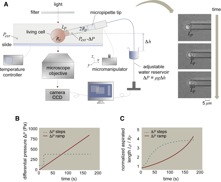
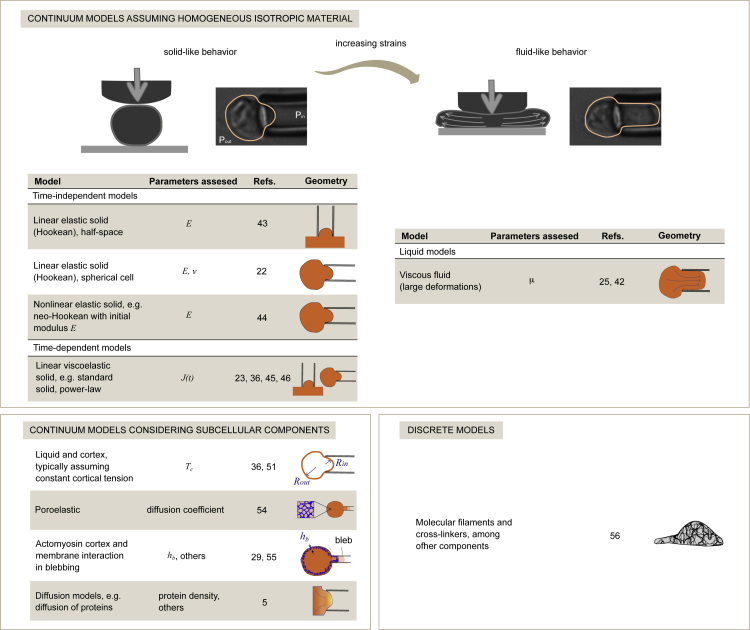
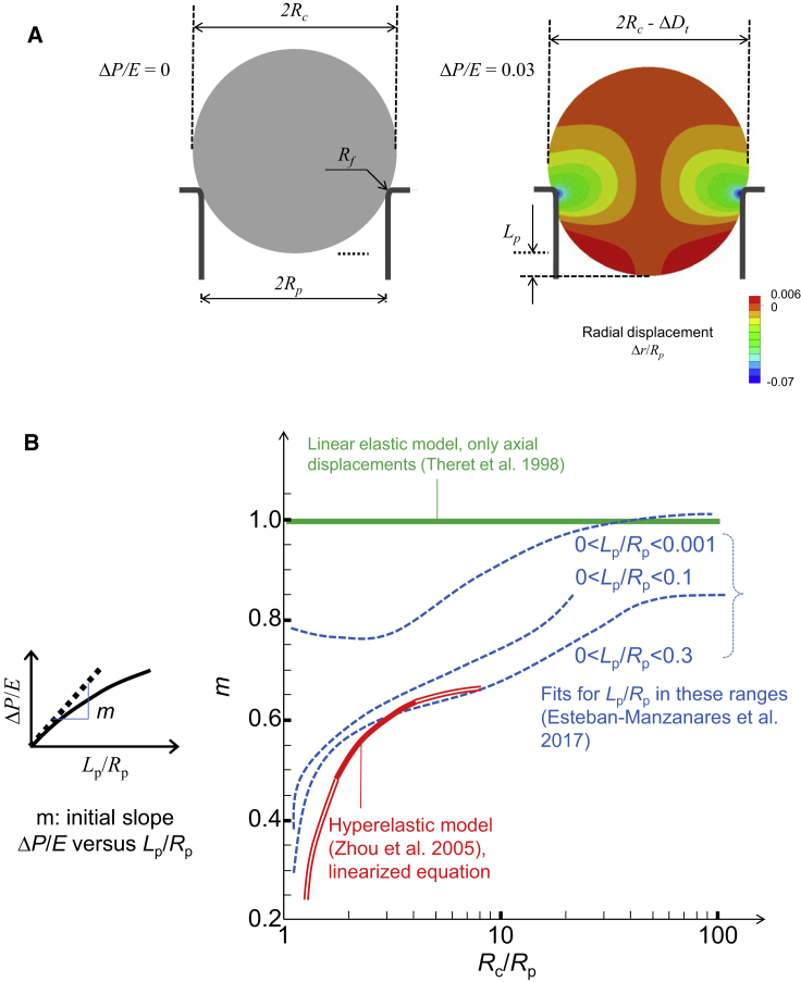
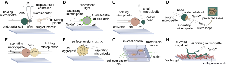
With five decades of sustained application, micropipette aspiration has enabled a wide range of biomechanical studies in the field of cell mechanics. Here, we provide an update on the use of the technique, with a focus on recent developments in the analysis of the experiments, innovative microaspiration-based approaches, and applications in a broad variety of cell types. We first recapitulate experimental variations of the technique. We then discuss analysis models focusing on important limitations of widely used biomechanical models, which underpin the urge to adopt the appropriate ones to avoid misleading conclusions. The possibilities of performing different studies on the same cell are also considered.


Figures

Figure 1
Figure 1
Scheme of the micropipette-aspiration experiments. (A) Experimental setup was used to simultaneously adjust the differential pressure ΔP, the temperature and the position of the micropipette, and the representative sequence of images of an experiment. (B) The graph shows two types of curves for the differential pressure ΔP applied over time: creep experiments at constant pressure (dashed line) and ramp experiments (solid line). (C) Plot of the aspirated length as a function of time for the two types of experiments. To see this figure in color, go online.
Figure 2
Figure 2
Models to analyze the mechanical properties of cells by micropipette aspiration. The figure summarizes continuum and discrete strategies for modeling the mechanical behavior of cells. To see this figure in color, go online.
Figure 3
Figure 3
Modeling the aspiration of a homogeneous, isotropic, and spherical cell. (A) Schemes showing geometrical parameters and radial displacements for a linear elastic sphere and a suction pressure ΔP/E = 0.03 (22). (B) Plot of the initial slope of the curves ΔP/E versus Lp/Rp as a function of Rc/Rp; slope was computed for Theret et al.’s model of a semi-infinite space (43), for Zhou et al.’s model of a hyperelastic sphere (44) (the linearized expression (30) is used, see (3), (4); the bold line represents the range originally studied by Zhou et al.), and for Esteban-Manzanares et al.’s model of a linear elastic sphere (22). In the latter case, the slope is represented for three ranges of fitting of the curve ΔP/E versus Lp/Rp. To see this figure in color, go online.
Figure 4
Figure 4
Schematic of micropipette-aspiration-based techniques. (A) Microindenter force tool to locally deform an attached cell. (B) Pipette aspiration was combined with confocal microscopy to relate protein expression (as measured by fluorescent microscopy intensity) and cell mechanical behavior. (C) Pipette-based force apparatus to measure cell pulling and pushing forces. (D) Cell-detachment technique was used to characterize adhesion forces. The resulting cell projected area as a function of time is related to the cell-substrate adhesion forces. (E) Dual-pipette aspiration was used to quantify intracellular forces. (F) Microaspiration device was used to map the surface tension of cell-aggregate interfaces. This mapping can be used to study morphogenesis and cell division. (G) Microfluidic high-throughput method was used to characterize single-cell mechanics. (H) Tip-pressure probe was used to assess the rate of growth of polarized cells. To see this figure in color, go online.

Similar articles

Cited by

References

    1. Tsai M.A., Frank R.S., Waugh R.E. Passive mechanical behavior of human neutrophils: power-law fluid. Biophys. J. 1993;65:2078–2088. - PMC - PubMed
    1. Keefer C.L., Desai J.P. Mechanical phenotyping of stem cells. Theriogenology. 2011;75:1426–1430. - PubMed
    1. Spagnol S.T., Lin W.C., Dahl K.N. Early passage dependence of mesenchymal stem cell mechanics influences cellular invasion and migration. Ann. Biomed. Eng. 2016;44:2123–2131. - PubMed
    1. van Helvert S., Storm C., Friedl P. Mechanoreciprocity in cell migration. Nat. Cell Biol. 2018;20:8–20. - PMC - PubMed
    1. Houk A.R., Jilkine A., Weiner O.D. Membrane tension maintains cell polarity by confining signals to the leading edge during neutrophil migration. Cell. 2012;148:175–188. - PMC - PubMed

Publication types

LinkOut - more resources