

Cognitive Control of Saccadic Selection and Inhibition from within the Core Cortical Saccadic Network
- PMID: 30683684
- PMCID: PMC6435832
- DOI: 10.1523/JNEUROSCI.1419-18.2018
Cognitive Control of Saccadic Selection and Inhibition from within the Core Cortical Saccadic Network
Abstract
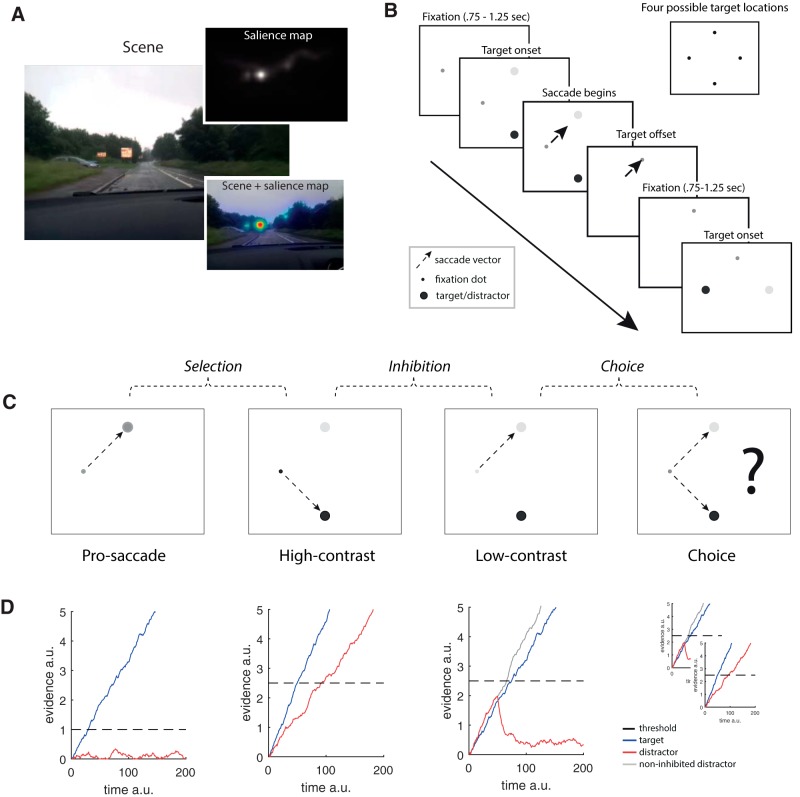
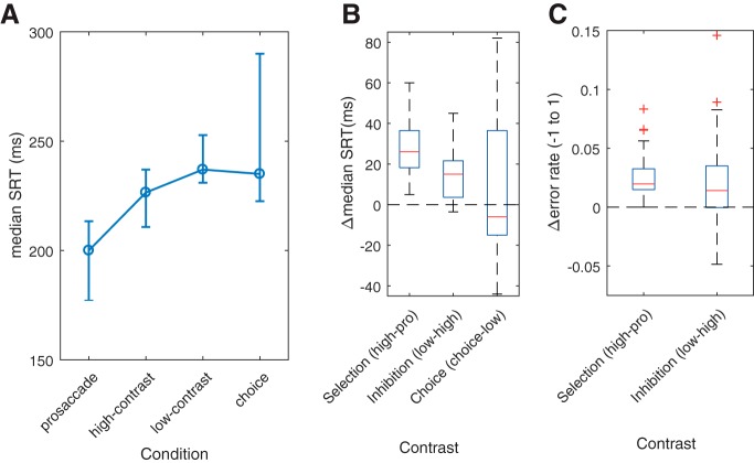
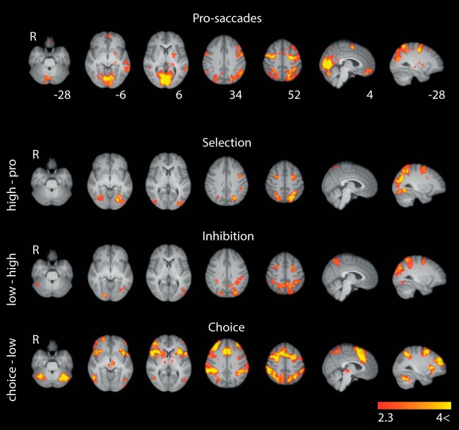
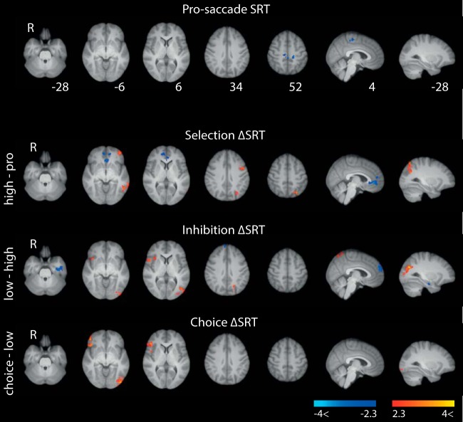
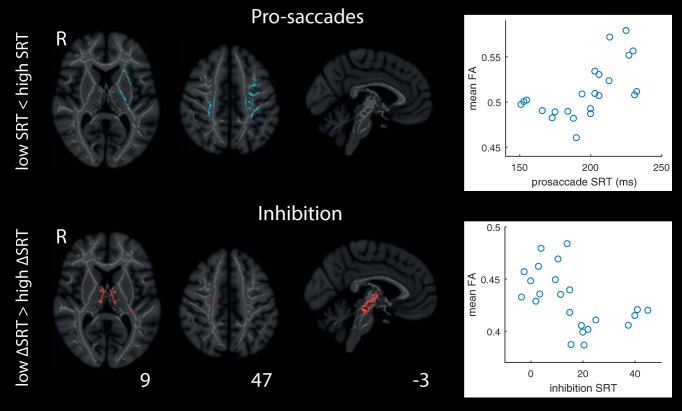
The ability to select the task-relevant stimulus for a saccadic eye movement, while inhibiting saccades to task-irrelevant stimuli, is crucial for active vision. Here, we present a novel saccade-contingent behavioral paradigm and investigate the neural basis of the central cognitive functions underpinning such behavior, saccade selection, saccade inhibition, and saccadic choice, in female and male human participants. The paradigm allows for exceptionally well-matched contrasts, with task demands formalized with stochastic accumulation-to-threshold models. Using fMRI, we replicated the core cortical eye-movement network for saccade generation (frontal eye fields, posterior parietal cortex, and higher-level visual areas). However, in contrast to previously published tasks, saccadic selection and inhibition recruited only this core network. Brain-behavior analyses further showed that inhibition efficiency may be underpinned by white-matter integrity of tracts between key saccade-generating regions, and that inhibition efficiency is associated with right inferior frontal gyrus engagement, potentially implementing general-purpose inhibition. The core network, however, was insufficient for saccadic choice, which recruited anterior regions commonly attributed to saccadic action selection, including dorsolateral prefrontal cortex and anterior cingulate cortex. Jointly, the results indicate that extra-saccadic activity observed for free choice, and in previously published tasks probing saccadic control, is likely due to increased load on higher-level cognitive processes, and not saccadic selection per se, which is achieved within the canonical cortical eye movement network.SIGNIFICANCE STATEMENT The ability to selectively attend to, and to not attend to, parts of the world is crucial for successful action. Mapping the neural substrate of the key cognitive functions underlying such behavior, saccade selection and inhibition, is a challenge. Canonical tasks, often preceding the cognitive neuroscience revolution by decennia, were not designed to isolate single cognitive functions, and result in extremely widespread brain activity. We developed a novel behavioral paradigm, which demonstrates the following: (1) the cognitive control of saccades is achieved within key cortical saccadic brain regions; (2) individual variability in control efficiency is related to white-matter connectivity between the same regions; and (3) widespread activity in canonical tasks is likely related to higher-level cognitive demands and not saccadic control.
Keywords: cognitive control; eye-tracking; fMRI; inhibition; saccades; selection.
Copyright © 2019 the authors 0270-6474/19/392497-12$15.00/0.
Figures
Similar articles
-
Task-based fMRI of a free-viewing visuo-saccadic network in the marmoset monkey.Neuroimage. 2019 Nov 15;202:116147. doi: 10.1016/j.neuroimage.2019.116147. Epub 2019 Aug 31. Neuroimage. 2019. PMID: 31479755
-
Developmental improvements in voluntary control of behavior: effect of preparation in the fronto-parietal network?Neuroimage. 2014 Sep;98:103-17. doi: 10.1016/j.neuroimage.2014.03.008. Epub 2014 Mar 15. Neuroimage. 2014. PMID: 24642280
-
Positron emission tomography study of voluntary saccadic eye movements and spatial working memory.J Neurophysiol. 1996 Jan;75(1):454-68. doi: 10.1152/jn.1996.75.1.454. J Neurophysiol. 1996. PMID: 8822570
-
The role of the parietal cortex in the neural processing of saccadic eye movements.Adv Neurol. 2003;93:141-57. Adv Neurol. 2003. PMID: 12894406 Review.
-
Neurophysiology and neuroanatomy of reflexive and volitional saccades: evidence from studies of humans.Brain Cogn. 2008 Dec;68(3):255-70. doi: 10.1016/j.bandc.2008.08.016. Epub 2008 Oct 5. Brain Cogn. 2008. PMID: 18835656 Free PMC article. Review.
Cited by
-
Brain Activity During Antisaccades to Faces in Adolescence.Cereb Cortex Commun. 2021 Sep 24;2(4):tgab057. doi: 10.1093/texcom/tgab057. eCollection 2021. Cereb Cortex Commun. 2021. PMID: 34806014 Free PMC article.
-
Neuronal activity in posterior parietal cortex area LIP is not sufficient for saccadic eye movement production.Front Integr Neurosci. 2023 Nov 24;17:1251431. doi: 10.3389/fnint.2023.1251431. eCollection 2023. Front Integr Neurosci. 2023. PMID: 38076390 Free PMC article.
-
Neural circuit disruptions of eye gaze processing in autism spectrum disorder and schizophrenia: An activation likelihood estimation meta-analysis.Schizophr Res. 2024 Feb;264:298-313. doi: 10.1016/j.schres.2023.12.003. Epub 2024 Jan 11. Schizophr Res. 2024. PMID: 38215566 Free PMC article.
-
Functional localization and categorization of intentional decisions in humans: A meta-analysis of brain imaging studies.Neuroimage. 2021 Nov 15;242:118468. doi: 10.1016/j.neuroimage.2021.118468. Epub 2021 Aug 11. Neuroimage. 2021. PMID: 34390878 Free PMC article.
-
Developmental maturation of causal signaling hubs in voluntary control of saccades and their functional controllability.Cereb Cortex. 2022 Oct 20;32(21):4746-4762. doi: 10.1093/cercor/bhab514. Cereb Cortex. 2022. PMID: 35094063 Free PMC article.
References
-
- Andersson JL, Jenkinson M, Smith S (2007) Non-linear registration, aka Spatial normalisation (FMRIB technical report TR07JA2) pp 1–21. Oxford: University of Oxford FMRIB Analysis Group.
Publication types
MeSH terms
Grants and funding
LinkOut - more resources
Full Text Sources