. 2019 Apr 3;39(14):2581-2605.
doi: 10.1523/JNEUROSCI.2234-18.2019. Epub 2019 Jan 25.

Presynaptic α2δ-2 Calcium Channel Subunits Regulate Postsynaptic GABAA Receptor Abundance and Axonal Wiring

Affiliations

Presynaptic α2δ-2 Calcium Channel Subunits Regulate Postsynaptic GABAA Receptor Abundance and Axonal Wiring

Stefanie Geisler et al. J Neurosci. .

Abstract

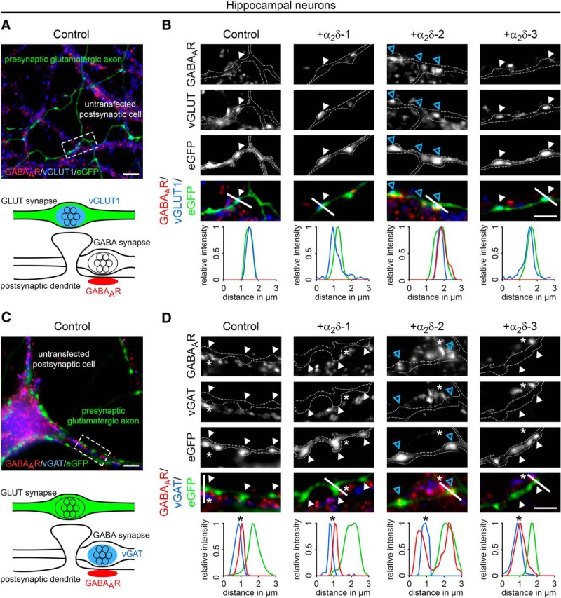
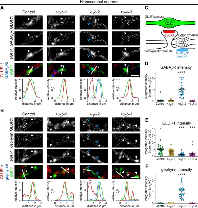
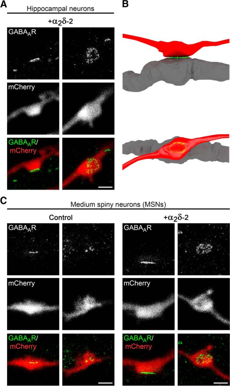
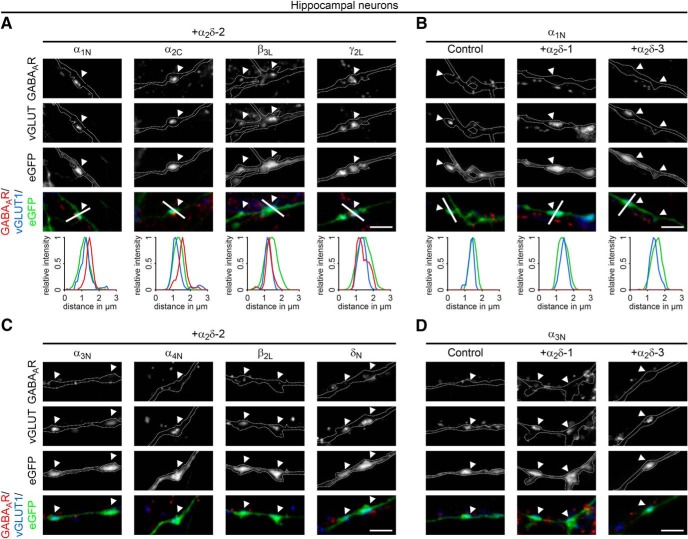
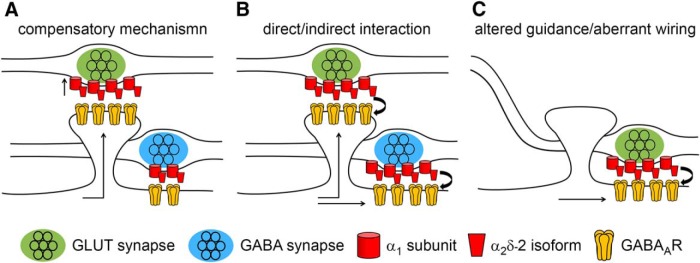
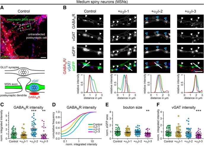
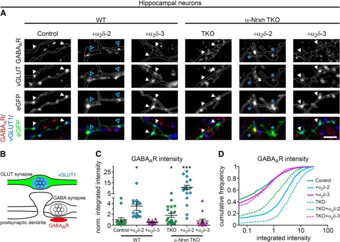
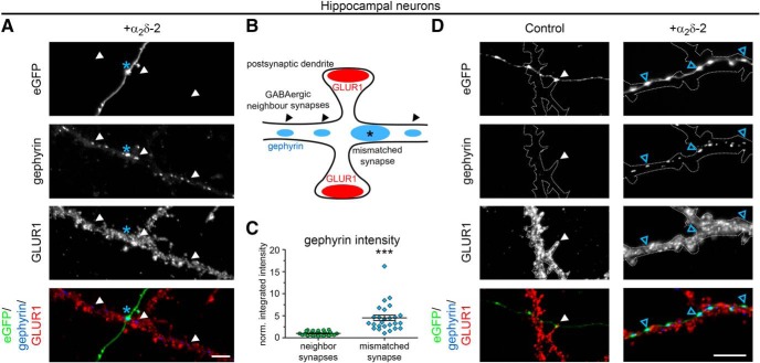
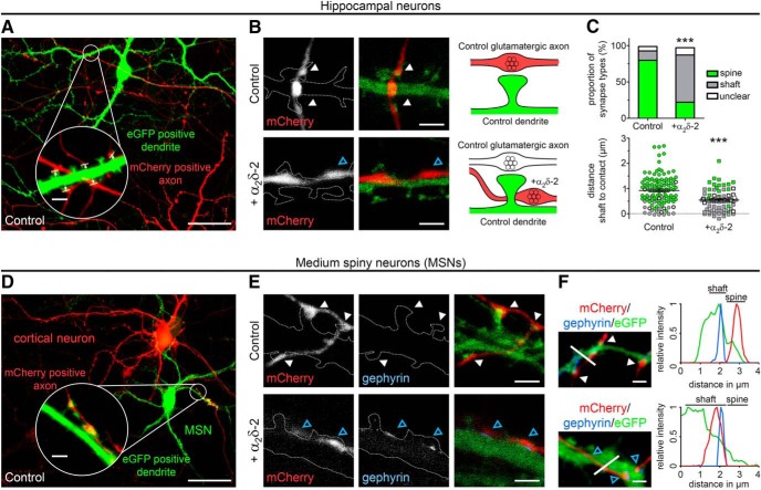
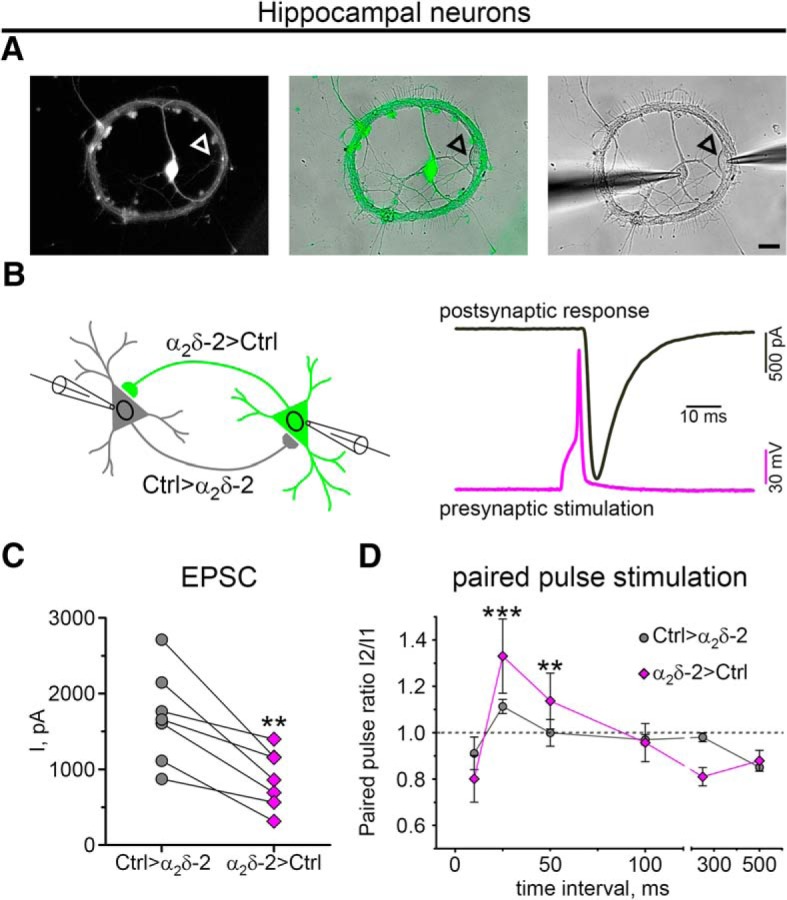
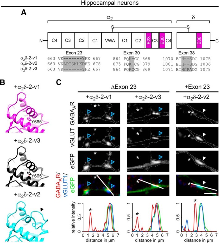
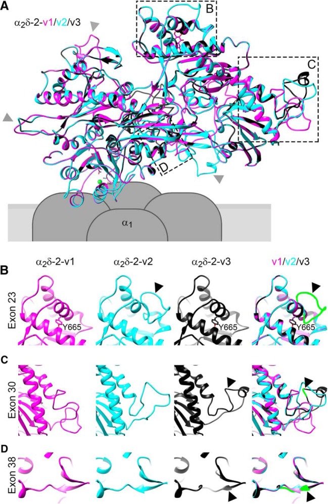
Presynaptic α2δ subunits of voltage-gated calcium channels regulate channel abundance and are involved in glutamatergic synapse formation. However, little is known about the specific functions of the individual α2δ isoforms and their role in GABAergic synapses. Using primary neuronal cultures of embryonic mice of both sexes, we here report that presynaptic overexpression of α2δ-2 in GABAergic synapses strongly increases clustering of postsynaptic GABAARs. Strikingly, presynaptic α2δ-2 exerts the same effect in glutamatergic synapses, leading to a mismatched localization of GABAARs. This mismatching is caused by an aberrant wiring of glutamatergic presynaptic boutons with GABAergic postsynaptic positions. The trans-synaptic effect of α2δ-2 is independent of the prototypical cell-adhesion molecules α-neurexins (α-Nrxns); however, α-Nrxns together with α2δ-2 can modulate postsynaptic GABAAR abundance. Finally, exclusion of the alternatively spliced exon 23 of α2δ-2 is essential for the trans-synaptic mechanism. The novel function of α2δ-2 identified here may explain how abnormal α2δ subunit expression can cause excitatory-inhibitory imbalance often associated with neuropsychiatric disorders.SIGNIFICANCE STATEMENT Voltage-gated calcium channels regulate important neuronal functions such as synaptic transmission. α2δ subunits modulate calcium channels and are emerging as regulators of brain connectivity. However, little is known about how individual α2δ subunits contribute to synapse specificity. Here, we show that presynaptic expression of a single α2δ variant can modulate synaptic connectivity and the localization of inhibitory postsynaptic receptors. Our findings provide basic insights into the development of specific synaptic connections between nerve cells and contribute to our understanding of normal nerve cell functions. Furthermore, the identified mechanism may explain how an altered expression of calcium channel subunits can result in aberrant neuronal wiring often associated with neuropsychiatric disorders such as autism or schizophrenia.

Keywords: auxiliary subunits; cacna2d; cultured hippocampal neurons; imaging; immunocytochemistry; voltage-gated calcium channels.


Figures

Figure 1.
Figure 1.
Presynaptic expression of α2δ-2 in cultured hippocampal neurons induces the formation of mismatched synapses. Representative immunofluorescence micrographs of cultured hippocampal neurons cotransfected with distinct α2δ subunits and soluble eGFP. Putative presynaptic en passent boutons (arrowheads) were identified as eGFP-filled axonal varicosities along dendrites of untransfected neurons (axons are outlined with dashed lines). A, B, Double labeling of transfected neurons (20–30 DIV) for vGLUT1 and the GABAAR β2/3 subunit. Colocalization of fluorescence signals was analyzed using line scans. A, In control boutons (eGFP only), potential glutamatergic synapses are positive for presynaptic vGLUT1 but negative for postsynaptic GABAAR (summarized in sketch). B, Presynaptic expression of α2δ-2 (blue arrowheads) induces postsynaptic GABAAR localization opposite transfected vGLUT1 positive nerve terminals. In contrast, postsynaptic GABAARs are not expressed opposite glutamatergic synapses expressing α2δ-1, α2δ-3, or eGFP only (control). C, D, Double labeling of transfected neurons (20–30 DIV) for vGAT and postsynaptic GABAAR. Colocalization of fluorescence signals was analyzed using line scans. C, In control boutons (eGFP only), potential glutamatergic synapses are negative for presynaptic vGAT and postsynaptic GABAAR (summarized in sketch). D, Transfected axonal varicosities were negative for vGAT in all conditions. Note the specific GABAAR labeling opposite vGAT-negative nerve terminals expressing α2δ-2 (blue arrowheads), which is in contrast to the colocalized fluorescence signals of vGAT and GABAAR in untransfected GABAergic neighboring synapses (asterisks). Representative images of two independent culture preparations are shown. Scale bars, 10 μm (A,C) and 3 μm (B,D).
Figure 2.
Figure 2.
Presynaptic expression of α2δ-2 alters the postsynaptic composition of glutamatergic synapses. A, B, Representative immunofluoresence micrographs of cultured hippocampal neurons (24–30 DIV) cotransfected with distinct α2δ subunits and soluble eGFP (control condition with eGFP alone is summarized in sketch, C). Colocalization of fluorescence signals was analyzed using line scans. A, Colabeling of the AMPA receptor subtype GLUR1 and the GABAAR. Presynaptic expression of α2δ-2 (blue arrowheads) induces postsynaptic GABAAR localization with a concomitant reduction of the GLUR1 fluorescence signal. In contrast, postsynaptic GABAARs are not expressed opposite putative glutamatergic synapses expressing α2δ-1, α2δ-3 or eGFP only (control). B, Colabeling of GLUR1 and gephyrin, the scaffolding protein of GABAergic synapses. Presynaptic expression of α2δ-2 (blue arrowheads) induces postsynaptic gephyrin localization with a concomitant reduction of the GLUR1 fluorescence signal. In contrast, postsynaptic gephyrin immunoreactivity is absent opposite putative glutamatergic synapses expressing α2δ-1, α2δ-3, or eGFP only (control). DF, Quantitative analysis of GABAAR (D), GLUR1 (E), and gephyrin (F) fluorescence intensities. Values were normalized to α2δ-2 (D, F) or eGFP only (control, E) fluorescence intensities within each culture preparation. Note the significant increase of postsynaptic GABAAR (D) and gephyrin (F) clusters opposite presynaptic boutons expressing α2δ-2. In contrast, integrated intensity of GLUR1 was significantly reduced opposite axonal varicosities transfected with α2δ-2 or α2δ-3. Values for individual cells (dots) and means (line) ± SEM are shown. Data are shown from three independent culture preparations; 11–25 (D), 22–30 (E), and 11–28 (F) cells were analyzed in each condition. Statistics: D, Kruskal–Wallis ANOVA with Dunn's post hoc analysis: H(4) = 51.6, p < 0.0001, post hoc: ****p < 0.0001 between α2δ-2 and all other conditions; E, ANOVA on log10-transformed data with Holm–Sidak post hoc analysis: F(3,100) = 18.6, p < 0.001, post hoc: ***p < 0.001 between control/α2δ-1 and α2δ-2/α2δ-3; F, Kruskal–Wallis ANOVA with Dunn's post hoc analysis: H(4) = 64.6, p < 0.0001, post hoc: ****p < 0.0001 between α2δ-2 and all other conditions. Asterisks in graphs indicate the significant difference compared with control. Scale bars, 3 μm.
Figure 3.
Figure 3.
GABAAR clusters are confined to the postsynaptic membrane. gSTED micrographs of cultured hippocampal (A) or MSNs (C) transfected with α2δ-2 and mCherry or mCherry only (control; 20–30 DIV). Transfected neurons (red, detected in confocal mode) were immunolabeled for the GABAAR (green, detected in gSTED mode). In all conditions, postsynaptic GABAAR clusters are closely opposed to mCherry-positive presynaptic boutons. B, 3D model showing that the GABAAR staining pattern depends on the orientation of the imaged synapse, which applies both for hippocampal as well as MSNs. Scale bar, 1 μm.
Figure 4.
Figure 4.
Presynaptic expression of α2δ-2 induces the recruitment of synaptic GABAAR subtypes. Representative immunofluorescence micrographs of cultured hippocampal neurons cotransfected with distinct α2δ subunits and soluble eGFP. Transfected permeabilized neurons (20–30 DIV) were double stained for vGLUT1 and different postsynaptic GABAAR subtypes. Colocalization of fluorescence signals within eGFP-filled axonal varicosities (arrowheads, axons are outlined with dashed lines) was analyzed using line scans. A, B, Immunofluorescence analysis identified intensely fluorescent clusters of the GABAAR subunits α1, α2, β3, and γ2 opposite α2δ-2 expressing glutamatergic (vGLUT1-positive) nerve terminals (A). In contrast, these postsynaptic GABAAR subtypes are absent opposite putative glutamatergic synapses expressing eGFP only (control), α2δ-1, or α2δ-3 (B, micrographs depict examples for α1). C, D, Labeling of the GABAAR subunits α3, α4, β2, and δ displayed weak and mainly extrasynaptic immunoreactivity in all conditions. Note that all GABAAR subtypes presented for α2δ-2 were also analyzed in hippocampal neurons expressing eGFP only, α2δ-1, and α2δ-3. Representative images of two independent cultures are shown. Scale bars, 3 μm.
Figure 5.
Figure 5.
Potential mechanisms explaining the observed mismatched synapse formation of glutamatergic nerve terminals expressing α2δ-2. A, Compensatory mechanism. Elevated expression of α2δ subunits increases presynaptic calcium channel abundance and current densities and thus glutamate release. Therefore, GABAARs could be recruited to the dendritic spine in an attempt to compensate for excessive excitatory synaptic activity. If this is the case, then GABAAR abundance at inhibitory synapses should not change upon α2δ-2 overexpression in GABAergic presynaptic terminals. B, α2δ-2 may be involved in trans-synaptically anchoring postsynaptic GABAARs. In this scenario, GABAAR abundance should be increased when α2δ-2 is overexpressed in both glutamatergic and GABAergic synapses. C, Trans-synaptic function of α2δ-2 could also induce aberrant axonal wiring by guiding glutamatergic axons to GABAergic postsynaptic locations positioned along dendritic shafts. This is in contrast to the normal situation in which glutamatergic synapses are generally formed on dendritic spines.
Figure 6.
Figure 6.
Striatum and cultured MSNs express three neuronal α2δ subunits. A, Absolute qRT-PCR analysis revealed a stable expression of α2δ-1, α2δ-2, and α2δ-3 in adult mouse striatum and monocultured MSNs (24–25 DIV). Although α2δ-3 was the dominant isoform in striatal tissue, mRNA levels for α2δ-2 and α2δ-3 were similarly abundant in cultured MSNs. Error bars indicate mean ± SEM. Data from three independent culture/tissue preparations are shown. B, β-galactosidase staining of sagittal cryosections of α2δ-3 knock-out mice carrying a LacZ cassette revealed intense labeling of striatum (Str), hippocampus (Hc), and olfactory tubercle (Ot). Lower expression was detected in the cortex (Cx), thalamus (Th), olfactory bulb (Ob), and parts of the cerebellum (Cb). C, Schematic representation of the epitope-tagged α2δ subunits depicting the position of the extracellular 2HA tag inserted downstream of the signal peptide (SP), cache domains (C1–C4), and VWA (van Willebrand factor type A). Cultured MSNs were transfected with HA-tagged α2δ subunits together with soluble eGFP and live labeled with an antibody against the HA epitope at 24 DIV. All α2δ isoforms are expressed at the surface of presynaptic boutons, which is also shown by line scan analysis of α2δ-1, α2δ-2, and α2δ-3 (red) in relation to synapsin (blue) and eGFP (green). The sketch summarizes the observed labeling patterns. Representative images of three (B) and one (C) independent preparation(s) are shown. Statistics: A, ANOVA on log10-transformed data with Holm–Sidak post hoc analysis: cultured MSNs: F(3,8) = 460, p < 0.001; post hoc: p < 0.001 between all α2δ subunits except α2δ-2 vs α2δ-3 (p = 0.34); striatum: F(3,8) = 891, p < 0.001; post hoc: p < 0.001 between all α2δ subunits except α2δ-1 vs α2δ-2 (p = 0.30). Scale bars, 2 mm (B) and 1 μm (C).
Figure 7.
Figure 7.
Presynaptic and postsynaptic differentiation of GABAergic MSNs. A, Schematic illustration of the coculture procedure: Cerebral hemispheres were dissected and stripped of meninges (1). Parts of the prefrontal cortex and striatum were dissected as shown (2). Striatal and cortical tissue was collected separately and dissociated using trypsin-EDTA and trituration (3). Cortical neurons were plated on poly-l-lysine-coated coverslips while striatal neurons were transfected with soluble eGFP (4). Subsequently, striatal neurons were plated onto cortical neurons in a ratio of 3:2 (5) and maintained above a glial feeder layer (6). B, Lentiviral infection or lipofection with soluble eGFP allowed discriminating MSNs from cortical neurons. CE, Double immunofluorescence of striatal–cortical cocultures (24–26 DIV) with presynaptic and postsynaptic markers for excitatory and inhibitory synapses. Neuronal morphology is outlined by eGFP. C, GABAergic synapses of transfected eGFP-positive MSNs showed immunoreactivity for vGAT, whereas vGLUT1 was absent. D, Axons of cortical neurons formed excitatory synapses on MSN spine heads (vGLUT1, white arrowheads), whereas GABAergic synapses were located on the dendritic shaft (vGAT, blue arrowhead). E, Labeling of PSD-95 in spine heads opposite synapsin-positive presynaptic terminals further indicated the presence of functional excitatory synapses on MSNs. F, Patch-clamp analysis of mIPSCs and mEPSCs in 14 DIV neurons confirmed the functionality of GABAergic and glutamatergic synapses. Representative micrographs of two independent cultures are shown. Scale bars, 50 μm (B), 3 μm (overview), and 1 μm (magnified selections; CE).
Figure 8.
Figure 8.
Presynaptic expression of α2δ-2 induces upregulation of postsynaptic GABAARs in MSNs. A, B, Representative immunofluorescence micrographs of cultured MSNs cotransfected with distinct α2δ subunits and soluble eGFP. Transfected neurons (21–28 DIV) were immunolabeled for vGAT and the GABAAR. Colocalization of fluorescence signals within eGFP-filled axonal varicosities (arrowheads, axons are outlined with dashed lines) was analyzed using line scans. A, GABAergic synapses transfected with eGFP only (control) show matched presynaptic vGAT and postsynaptic GABAAR immunoreactivity (summarized in sketch). B, Similar to control, postsynaptic GABAARs were localized opposite vGAT-positive presynaptic terminals expressing individual α2δ isoforms (see also colocalization in line scans). Most importantly, GABAAR clusters were larger and more intense opposite synaptic boutons expressing α2δ-2 (blue arrowheads). CF, Quantitative analysis of GABAAR fluorescence intensity (C), cumulative frequency distribution of GABAAR fluorescence intensity (D), bouton size (E), and vGAT fluorescence intensity (F). Values for individual cells (dots) and means (lines) ± SEM are shown. Values were normalized to the control within each culture preparation. Data from four independent culture preparations and 34–42 cells were analyzed in each condition. Statistics: ANOVA on log10-transformed data with Holm–Sidak post hoc analysis: C, F(3,152) = 17.6, p < 0.001; post hoc: ***p < 0.001 between α2δ-2 and control, ***p < 0.001 between α2δ-2 and α2δ-1/α2δ-3, **p = 0.004 between control and α2δ-3, p = 0.2 between control and α2δ-1; E, F(3,152) = 5.1, p < 0.01; post hoc: **p < 0.01 between α2δ-3 and control/α2δ-2, p = 0.08 between α2δ-3 and α2δ-1; F, F(3,152) = 8.0, p < 0.001; post hoc: ***p < 0.001 between α2δ-3 and control, **p < 0.01 between α2δ-3 and α2δ-1/α2δ-2. Asterisks in graphs indicate the significant difference compared with control. Scale bars, 10 μm (A) and 3 μm (B).
Figure 9.
Figure 9.
α-Nrxn modulates the effect of α2δ-2 on GABAARs. Representative immunofluorescence micrographs of cultured hippocampal neurons (20–25 DIV) prepared from WT or α-Nrxn TKO mice. A, B, In control boutons (eGFP only), potential glutamatergic synapses are positive for presynaptic vGLUT1 but negative for postsynaptic GABAAR staining (summarized in sketch, B). Consistent with our initial observation (see Fig. 1), presynaptic expression of α2δ-2 (blue arrowheads) induces postsynaptic GABAAR localization opposite vGLUT1-positive nerve terminals in WT neurons. Most importantly, in α-Nrxn TKO synapses, the expression of α2δ-2 strongly induced postsynaptic GABAAR localization (blue asterisks). C, Postsynaptic GABAARs clusters (integrated intensities) are significantly increased opposite glutamatergic nerve terminals expressing α2δ-2 compared with control (eGFP only). This effect is much stronger in α-Nrxn TKO synapses compared with WT neurons (3-fold higher GABAAR fluorescence intensity). D, Cumulative frequency distribution further reveals that the vast majority of glutamatergic boutons are positive for postsynaptic GABAARs in WT+α2δ-2 (88%) and TKO+α2δ-2 (97%) and that the population is shifted toward larger and more intense clusters. However, this analysis also demonstrates that mismatched GABAARs already form opposite α-Nrxn TKO synapses, although at a lower overall intensity. Values for individual cells (dots) and means (lines) ± SEM are shown. Values were normalized to the WT control within each culture preparation. Data from two independent culture preparations and 12–19 cells were analyzed within each condition. Statistics: C, ANOVA on log10-transformed data with Holm–Sidak post hoc analysis: genotype: F(1,86) = 5.3, p = 0.023; transfection: F(2,86) = 49.0, p < 0.001; genotype × transfection: F(2,86) = 3.7, p = 0.03; post hoc: *p < 0.05, ***p < 0.001. Asterisks in graphs indicate the significant difference compared with control. Scale bar, 3 μm.
Figure 10.
Figure 10.
Mismatched synapses show characteristics of GABAergic shaft synapses. A, D, Representative immunofluorescence micrographs of cultured hippocampal neurons cotransfected with α2δ-2 and soluble eGFP. Colabeling of transfected neurons (24 DIV) for postsynaptic GLUR1 and gephyrin is shown. A, Presynaptic expression of α2δ-2 induces a strong upregulation of postsynaptic gephyrin (asterisk) compared with neighboring endogenous GABAergic synapses situated on the same dendrite (arrowheads). Although GLUR1 expression was reduced in mismatched synapses (*; see Fig. 2), it was absent in endogenous GABAergic synapses (arrowheads, see sketch in B). C, Quantitative analysis of gephyrin fluorescence intensities in GABAergic neighboring and mismatched synapses. Dots represent values for individual boutons (mismatched synapses) and means of 10 endogenous clusters measured per image. Results were normalized to endogenous gephyrin intensities of neighboring synapses within each culture preparation. Data from three independent culture preparations are shown. Statistics: t test on log10-transformed data: t(50) = 9.5; ***p < 0.001. D, Left, Control boutons transfected with eGFP only form glutamatergic synapses on dendritic spines (gephyrin negative, GLUR1 positive; white arrowheads). Right, Expression of α2δ-2 in putative glutamatergic boutons induces the formation of mismatched synapses (gephyrin-positive, GLUR1-negative) along the shaft of the dendrite (blue arrowheads). Dendritic morphology of untransfected neurons was outlined according to GLUR1 labeling. Scale bars, 5 μm (A) and 4 μm (B).
Figure 11.
Figure 11.
Presynaptic α2δ-2 induces the aberrant wiring of glutamatergic axons to dendritic shafts. A, D, To analyze the position of α2δ-2-induced mismatched synapses on postsynaptic dendrites of excitatory (A, hippocampal neurons) and inhibitory neurons (D, MSNs), presynaptic (control or α2δ-2) and postsynaptic neurons were labeled with mCherry (red) and eGFP (green), respectively (for details, see Materials and Methods). The magnified insets in A and D demonstrate the close association of presynaptic axonal varicosities (mCherry) with postsynaptic dendritic spines (eGFP), as expected for excitatory spine synapses. Using gSTED microscopy, the distance from the dendritic shaft to the contact zone of each synaptic bouton was measured (A, arrows and lines in the magnified inset). B, E, gSTED microscopy confirms the preferential location of excitatory synapses on dendritic spines of both glutamatergic or GABAergic neurons (white arrowheads and sketch in B) and, most importantly, suggests an aberrant wiring of putative glutamatergic axons expressing α2δ-2 with postsynaptic sites along dendritic shafts (blue arrowheads and sketch in B), as typically observed for GABAergic synapses. C, Corresponding postsynaptic location of each contacting bouton was categorized as spine (green), shaft (gray), or as unclear (white; top) and the distance from the dendritic shaft to the contact point was measured (bottom). Although the vast majority of synaptic contact points in the control condition are located on dendritic spines (80%), presynaptic expression of α2δ-2 shifted the preferential contact points to dendritic shafts (65%, top). This is further confirmed by a significantly decreased contact point to shaft distance of α2δ-2-expressing compared with control synapses. Values for individual boutons (dots) and means (lines) ± SEM are shown. Dot colors (bottom) show the respective categorized synapse position (spine, green; shaft, gray; and unclear, white) presented in the top. Data from two independent culture preparations and 118 (control) and 75 (+α2δ-2) boutons were analyzed. Statistics: C, top: χ2 test: χ2(2) = 63.4, ***p < 0.001; bottom: Mann–Whitney U test: ***p < 0.001. E, F, gSTED microscopy (E) and high-resolution fluorescence microscopy (F) revealed that, similar to hippocampal neurons, presynaptic expression of α2δ-2 in cortical neurons shifted the preferential contact points to dendritic shafts of GABAergic MSNs, as indicated by gephyrin labeling and the dendritic position (blue arrowheads and line scan in F). Representative micrographs of two independent cultures are shown. Scale bars, 50 μm (overview), 3 μm (insets, A, D), and 2 μm (B, E, F).
Figure 12.
Figure 12.
Presynaptic expression of α2δ-2 affects synaptic transmission in aberrantly wired synapses. AC, Experimental paradigm to study the functional consequences of presynaptic α2δ-2 overexpression in mismatched glutamatergic synapses of cultured hippocampal neurons. A, Neurons were cultured in pairs of two synaptically connected cells in which one represents an untransfected control neuron (arrowhead) and the other an α2δ-2-overexpressing neuron (eGFP labeled). B, Whole-cell patch-clamp recordings of excitatory synaptic transmission (13–18 DIV). An action potential in neuron 1 elicited by a given AP waveform recorded from WT hippocampal neurons caused eEPSCs in neuron 2. Synaptic transmission was analyzed in both directions of synaptically connected pairs. Therefore, putative glutamatergic spine synapses (control innervating α2δ-2-overexpressing neuron) and putative mismatched synapses (α2δ-2 overexpressing innervating control neuron) could be directly compared within the same pair. C, D, Quantitative analysis of mean eEPSC peak amplitudes (C) and paired-pulse response ratios (PPRs) as a measure for synaptic plasticity (D). C, eEPSCs were strongly reduced (48%) in mismatched synapses. D, Dashed line shows the boundary between paired-pulse depression (PPR < 1) and facilitation (PPR > 1). Note the slight significant increase in facilitation in mismatched synapses at 25 and 50 ms compared with the initial 10 ms interval. Values for individual pairs (dots) and means (lines) ± SEM are shown. Data from three independent culture preparations from seven (C) and six (D) pairs were analyzed. Statistics: C, paired t test: t(6) = 4.6; **p < 0.01; D, two-way repeated-measures ANOVA: condition: F(1,45) = 0.02, p = 0.9; interval: F(5,45) = 11.1, p < 0.001; condition × interval: F(5,45) = 2.7, p = 0.03; Holm–Sidak post hoc analysis: **p = 0.004, ***p < 0.001 compared with 10 ms within α2δ-2 to control. Scale bar, 20 μm.
Figure 13.
Figure 13.
Lack of exon 23 in α2δ-2 splice variants mediates the trans-synaptic effect on GABAARs. A, Schematic overview illustrating the approximate positions of three alternatively spliced regions of α2δ-2. Sequence alignment between α2δ-2-v1, the original construct used in this study, and two additional α2δ-2 variants (v2, v3) reveals alternative splicing of exons 23, 30, and 38. B, Using homology modeling, we tested the potential consequences of alternative splicing on the structure prediction based on the high-resolution structure of α2δ-1 (PDB code: 5GJV; Wu et al., 2016; see Fig. 14). Inclusion of exon 23 in α2δ-2-v2 suggested the formation of an extra loop (arrowhead) leading to the disruption of an α-helix present in α2δ-2-v1 and α2δ-2-v3. C, Representative immunofluorescence micrographs of cultured hippocampal neurons (21–26 DIV) cotransfected with distinct α2δ-2 splice variants and soluble eGFP. Presynaptic expression of either α2δ-2-v1 (original construct) or α2δ-2-v3, which both lack exon 23, robustly induces a mismatched GABAAR localization opposite vGLUT1-positive nerve terminals (blue arrowheads). In contrast, presynaptic expression α2δ-2-v2, which contains exon 23, failed to induce mismatched synapse formation (white arrowheads, line scans). Asterisks mark endogenous GABAAR clusters from untransfected neighboring synapses not colocalizing with presynaptic vGLUT1. Representative images of two independent cultures are shown. Scale bar, 3 μm.
Figure 14.
Figure 14.
Splicing sites of α2δ-2 and implications on secondary structure. A, Using homology modeling, we tested the potential consequences of alternative splicing on the structure prediction of three distinct α2δ-2 splice variants (v1–v3) based on the high-resolution structure of α2δ-1 (PDB code: 5GJV; Wu et al., 2016). Although some of the longer loops generally seemed to be quite flexible and differently orientated in all three models (gray arrows), alternative splicing of exons 23, 30, and 38 resulted in differences between secondary structure elements in distinct variants (boxed regions in BD). B, Higher magnification showing that the inclusion of exon 23 in α2δ-2-v2 suggests the formation of an extra loop (arrowhead and green selection in overlay), leading to the disruption of an α-helix present in α2δ-2-v1 and α2δ-2-v3 (see also Fig. 13). C, Structure modeling further proposes that the insertion of three base pairs in exon 30 of α2δ-2-v3 causes the formation of a short α-helix in the depicted loop, which is absent in α2δ-2-v1 and α2δ-2-v2. D, Moreover, models implicate that alternative splicing of exon 38 affects the length of a β sheet at this position, which is longer in α2δ-2-v1 and α2δ-2-v2.

References

    1. Anderson TR, Shah PA, Benson DL (2004) Maturation of glutamatergic and GABAergic synapse composition in hippocampal neurons. Neuropharmacology 47:694–705. 10.1016/j.neuropharm.2004.07.023 - DOI - PubMed
    1. Aoto J, Martinelli DC, Malenka RC, Tabuchi K, Südhof TC (2013) Presynaptic neurexin-3 alternative splicing trans-synaptically controls postsynaptic AMPA receptor trafficking. Cell 154:75–88. 10.1016/j.cell.2013.05.060 - DOI - PMC - PubMed
    1. Aoto J, Földy C, Ilcus SM, Tabuchi K, Südhof TC (2015) Distinct circuit-dependent functions of presynaptic neurexin-3 at GABAergic and glutamatergic synapses. Nat Neurosci 18:997–1007. 10.1038/nn.4037 - DOI - PMC - PubMed
    1. Arama J, Abitbol K, Goffin D, Fuchs C, Sihra TS, Thomson AM, Jovanovic JN (2015) GABAA receptor activity shapes the formation of inhibitory synapses between developing medium spiny neurons. Front Cell Neurosci 9:290. 10.3389/fncel.2015.00290 - DOI - PMC - PubMed
    1. Ba-Abbad R, Arno G, Carss K, Stirrups K, Penkett CJ, Moore AT, Michaelides M, Raymond FL, Webster AR, Holder GE (2016) Mutations in CACNA2D4 cause distinctive retinal dysfunction in humans. Ophthalmology 123:668–671.e2. 10.1016/j.ophtha.2015.09.045 - DOI - PubMed

Publication types

MeSH terms

Associated data