. 2019 Mar 22;294(12):4498-4510.
doi: 10.1074/jbc.RA118.007260. Epub 2019 Jan 25.

Influence of the N-terminal segment and the PHY-tongue element on light-regulation in bacteriophytochromes

Affiliations

Influence of the N-terminal segment and the PHY-tongue element on light-regulation in bacteriophytochromes

Geoffrey Gourinchas et al. J Biol Chem. .

Abstract

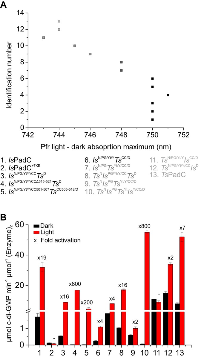
Photoreceptors enable the integration of ambient light stimuli to trigger lifestyle adaptations via modulation of central metabolite levels involved in diverse regulatory processes. Red light-sensing bacteriophytochromes are attractive targets for the development of innovative optogenetic tools because of their natural modularity of coupling with diverse functionalities and the natural availability of the light-absorbing biliverdin chromophore in animal tissues. However, a rational design of such tools is complicated by the poor understanding of molecular mechanisms of light signal transduction over long distances-from the site of photon absorption to the active site of downstream enzymatic effectors. Here we show how swapping structural elements between two bacteriophytochrome homologs provides additional insight into light signal integration and effector regulation, involving a fine-tuned interplay of important structural elements of the sensor, as well as the sensor-effector linker. Facilitated by the availability of structural information of inhibited and activated full-length structures of one of the two homologs (Idiomarina species A28L phytochrome-activated diguanylyl cyclase (IsPadC)) and characteristic differences in photoresponses of the two homologs, we identify an important cross-talk between the N-terminal segment, containing the covalent attachment site of the chromophore, and the PHY-tongue region. Moreover, we highlight how these elements influence the dynamic range of photoactivation and how activation can be improved to light/dark ratios of ∼800-fold by reducing basal dark-state activities at the same time as increasing conversion in the light state. This will enable future optimization of optogenetic tools aiming at a direct allosteric regulation of enzymatic effectors.

Keywords: GGDEF; bilin; cyclic di-GMP (c-di-GMP); diguanylate cyclase; photobiology; photoreceptor; phytochrome; protein engineering; signal transduction; ultraviolet-visible spectroscopy (UV-visible spectroscopy).


Conflict of interest statement

The authors declare that they have no conflicts of interest with the contents of this article

Figures

Figure 1.
Figure 1.
Conservation of residues between BV surrounding structural elements in IsPadC and TsPadC. A, cartoon representation of the dark-adapted full-length structure of IsPadC (15). Subdomains are colored as follow: NTS, black; PAS, purple; GAF, blue; PHY, green; coiled-coil linker, orange; and DGC, red. B, sequence alignment generated using Clustal Omega (56) with standard settings and colored by residue conservation using Jalview (57). The alignment is represented for important structural features of the IsPadC and TsPadC photosensory modules and the coiled-coil linker regions. C, close-up view of the NTS interactions with the GAF domain and the PHY-tongue element in the Pr state of IsPadC. ZZZssa BV and important residues involved in the interactions are highlighted in stick representation. D, close-up view of the typical phytochrome GAF knot lasso of IsPadC. Important conserved residues involved in the stabilization of the NTS element are highlighted in stick representation.
Figure 2.
Figure 2.
Comparison of UV-visible absorption spectra of IsPadC and TsPadC. A, superposition of IsPadC (solid lines) and TsPadC (dashed lines) UV-visible spectra, normalized based on the IsPadC Soret-band maximum. Difference spectra of illuminated and dark-state spectrum are represented in blue. B, denaturation in methanol with 0.1% TCA of IsPadC and TsPadC under both dark and illuminated conditions. The same denaturation of DrBphP is shown as a reference of complete ZZEssa formation upon illumination. C, superposition of room temperature (20 °C) and low temperature (2 °C) absorption spectra of IsPadC (solid lines) and TsPadC (dashed lines). Spectra are scaled together based on the Soret peak absorbance.
Figure 3.
Figure 3.
Overview of generated constructs. A, schematic representation of the domain arrangement of naturally occurring constructs and different synthetic chimeras. Subdomain representations of IsPadC and TsPadC are colored in black and light gray, respectively. The coiled-coil regions are colored in orange and light orange for IsPadC and TsPadC, respectively. Residue numbers of the fusion points are indicated. Names of individual subdomains are colored according to Fig. 1A. B, in vivo screening of DGC activity in Congo Red–based assays under dark, as well as constant red light illumination. Bacterial colonies expressing active DGCs are colored red because of the Congo Red dye being complexed by exopolysaccharides that are produced upon c-di-GMP formation (58). Isolated bacterial colonies are shown in the order of the construct presented in A. The full screening plates with all colonies together are available in Fig. S2.
Figure 4.
Figure 4.
UV-visible absorption spectra of IsPadC and TsPadC as well as synthetic chimeras. The dark-state spectrum is colored black, whereas the illuminated steady-state spectrum is colored red. The difference spectrum between the illuminated and dark states is colored blue, and the maxima of positive and negative differences are indicated. On the left side of each spectrum, a schematic representation of the synthetic fusion is shown where black domains derive from IsPadC, and light gray domains derive from TsPadC. For the fusions IsN/PGTsYt/Y/CC/D, TsNIsPGTsYt/Y/CC/D, and TsNIsPGTsYtIsY/CC/D, UV-visible spectra of the illuminated state in green were also recorded at 2 °C to minimize the effect of spontaneous thermal reversion of Pfr states and to reduce shunt reaction pathways from photocycle intermediates. The difference spectrum between the illuminated and dark state at 2 °C is colored light blue.
Figure 5.
Figure 5.
Regulation capacity of DGC activity. A, maxima of the far-red light absorption derived from the difference between illuminated and dark state spectra of all constructs. B, comparison of apparent turnover rate constants between dark and light conditions at 200 μm GTP. The fold activation is indicated above the columns. Initial rates are quantified from experimental triplicates of three time points, and the sample standard deviation of individual points contributed to the error estimation of the linear fit that is used to calculate the initial rate of product formation. The S.E. of the estimate from the linear regression is used as an error indicator.

Similar articles

Cited by

References

    1. Heintzen C. (2012) Plant and fungal photopigments. Wiley Interdiscip. Rev. Membr. Transp. Signal. 1, 411–432 10.1002/wmts.36 - DOI
    1. Gomelsky M., and Hoff W. D. (2011) Light helps bacteria make important lifestyle decisions. Trends Microbiol. 19, 441–448 10.1016/j.tim.2011.05.002 - DOI - PubMed
    1. Losi A., and Gärtner W. (2017) Solving blue light riddles: new lessons from flavin-binding LOV photoreceptors. Photochem. Photobiol. 93, 141–158 10.1111/php.12674 - DOI - PubMed
    1. Rockwell N. C., Su Y.-S., and Lagarias J. C. (2006) Phytochrome structure and signaling mechanisms. Annu. Rev. Plant Biol. 57, 837–858 10.1146/annurev.arplant.56.032604.144208 - DOI - PMC - PubMed
    1. Duanmu D., Bachy C., Sudek S., Wong C.-H., Jiménez V., Rockwell N. C., Martin S. S., Ngan C. Y., Reistetter E. N., van Baren M. J., Price D. C., Wei C. L., Reyes-Prieto A., Lagarias J. C., and Worden A. Z. (2014) Marine algae and land plants share conserved phytochrome signaling systems. Proc. Natl. Acad. Sci. U.S.A. 111, 15827–15832 10.1073/pnas.1416751111 - DOI - PMC - PubMed

Publication types

MeSH terms

LinkOut - more resources