. 2019 Feb 12;116(7):2603-2611.
doi: 10.1073/pnas.1818313116. Epub 2019 Jan 25.

Cells exhibiting strong p16INK4a promoter activation in vivo display features of senescence

Affiliations

Cells exhibiting strong p16INK4a promoter activation in vivo display features of senescence

Jie-Yu Liu et al. Proc Natl Acad Sci U S A. .

Abstract

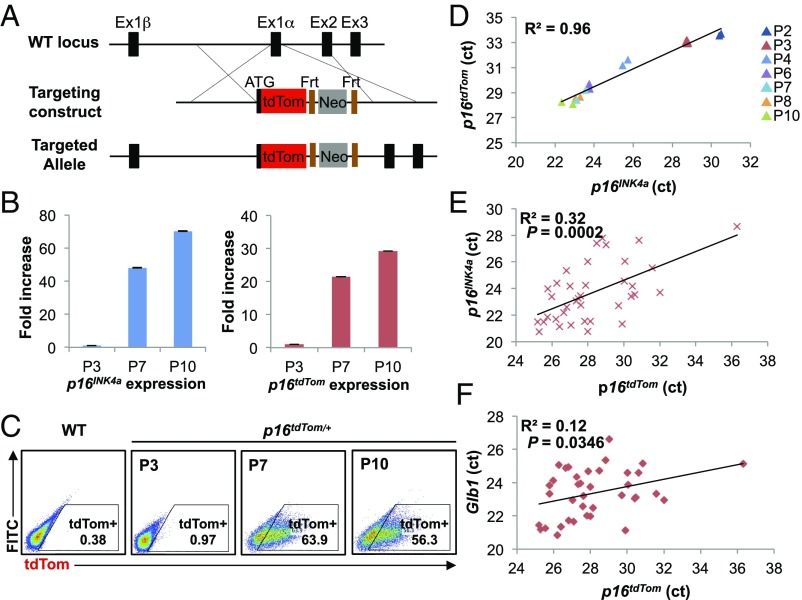
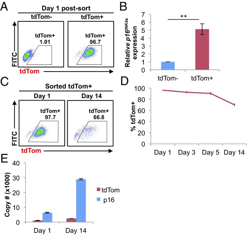
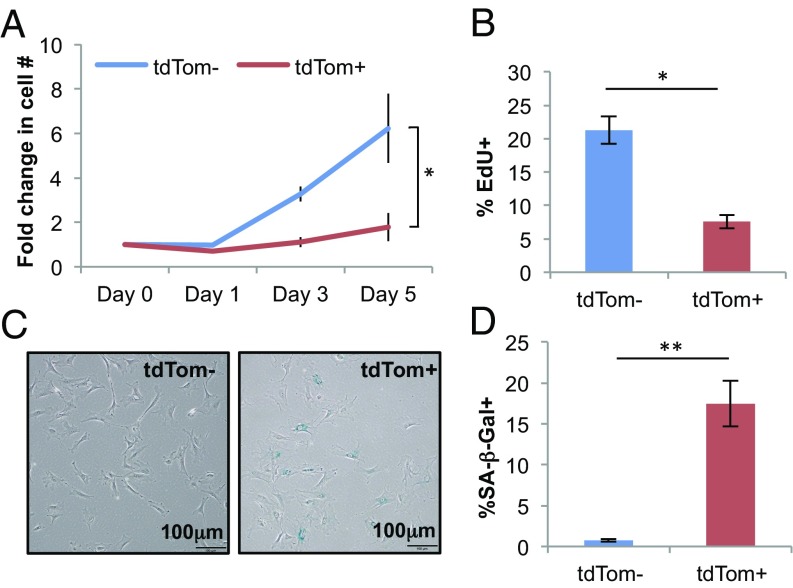
The activation of cellular senescence throughout the lifespan promotes tumor suppression, whereas the persistence of senescent cells contributes to aspects of aging. This theory has been limited, however, by an inability to identify and isolate individual senescent cells within an intact organism. Toward that end, we generated a murine reporter strain by "knocking-in" a fluorochrome, tandem-dimer Tomato (tdTom), into exon 1α of the p16INK4a locus. We used this allele (p16tdTom ) for the enumeration, isolation, and characterization of individual p16INK4a -expressing cells (tdTom+). The half-life of the knocked-in transcript was shorter than that of the endogenous p16INK4a mRNA, and therefore reporter expression better correlated with p16INK4a promoter activation than p16INK4a transcript abundance. The frequency of tdTom+ cells increased with serial passage in cultured murine embryo fibroblasts from p16tdTom/+ mice. In adult mice, tdTom+ cells could be readily detected at low frequency in many tissues, and the frequency of these cells increased with aging. Using an in vivo model of peritoneal inflammation, we compared the phenotype of cells with or without activation of p16INK4a and found that tdTom+ macrophages exhibited some features of senescence, including reduced proliferation, senescence-associated β-galactosidase (SA-β-gal) activation, and increased mRNA expression of a subset of transcripts encoding factors involved in SA-secretory phenotype (SASP). These results indicate that cells harboring activation of the p16INK4a promoter accumulate with aging and inflammation in vivo, and display characteristics of senescence.

Keywords: aging; cdkn2a; senescence.


Conflict of interest statement

The authors declare no conflict of interest.

Figures

Fig. 1.
Fig. 1.
Design and validation of the p16tdTom allele. (A) Schematic of the p16tdTom knockin targeting strategy. Frt, flippase recognition site; Neo, neomycin resistance gene. (BD) Induction of p16INK4a and tdTom expression in p16tdTom/+ MEFs over serial passage. P3, passage 3; P7, passage 7; P10, passage 10. mRNA expression of p16INK4a and tdTom by qRT-PCR. Fold-increase was calculated with respect to the mRNA levels at P3. Data shown correspond to three biological replicates. Error bars represent SEM (B). Representative flow cytometric (FACS) analysis of MEFs at indicated passage number (C). Correlation of p16INK4a and p16tdTom mRNA expression shown in the normalized threshold cycle (ct) value. Passage number is presented by different colors. Linear regression was used to calculate the coefficient of determination (R2) (D). (E and F) Linear correlations between p16INK4a, p16tdTom, and Glb1 mRNA expression on single-cell levels. Expression levels are shown in the comparative threshold cycle (ct) values. Each dot represents a single cell.
Fig. 2.
Fig. 2.
Promoter activity of the p16tdTom allele. (A) Representative FACS analysis of tdTom and tdTom+ populations at day 1 postsort. (B) mRNA expression of p16INK4a by qRT-PCR. Fold-difference was calculated with respect to the mRNA levels in tdTom MEFs (**P < 0.01). (C) Representative FACS analysis of the cultured tdTom+ MEFs at indicated time points after cell sorting. (D) Frequency of tdTom+ cells in the tdTom+ cultures at indicated time points after cell sorting. (E) Absolute copy number of p16INK4a and tdTom in cultured tdTom+ MEFs by qRT-PCR at indicated time points after cell sorting. Throughout, error bars represent SEM.
Fig. 3.
Fig. 3.
p16INK4a-activated cultured MEFs exhibit senescence phenotypes. (A) Growth-curve analysis of tdTom and tdTom+ populations. Fold-increase was calculated with respect to the cell number at day 0. (B) Quantification of EdU+ cells by immunofluorescence staining. (C) Representative image of SA-β-gal staining. (D) Quantification of SA-β-gal+ cells in C. Throughout, error bars represent SEM. The statistical significance of differences was assessed using paired two-tailed Student’s t tests (*P < 0.05, **P < 0.01).
Fig. 4.
Fig. 4.
Age-dependent increase in the frequency of p16INK4a-activated cells in different tissues. (AD) Representative FACS analysis of CD3+ (T cells), B220+ and Mac-1+ (myeloid cells) populations from peripheral blood (PB) (A), cartilages (B), fat progenitor cells (Sca1+CD34+) from IAT (C), and pancreatic islets (D). Tissues were harvested from young (8- to 12-wk-old) and old (100- to 120-wk-old) p16tdTom/+ mice. (E) Quantification of tdTom+ cells from the indicated tissues. Error bars represent SEM (n = 3–4 per group). The statistical significance of differences was assessed using unpaired two-tailed Student’s t tests (*P < 0.05).
Fig. 5.
Fig. 5.
Reduced proliferation and high SA-β-gal activity of p16INK4a-activated peritoneal macrophages. (A) Bioluminescence imaging of p16LUC/+ mice following intraperitoneal injection with empty (control, CNTL) or quiescent human NDF-embedded alginate beads. Representative image was acquired 21 d after bead injection. (B) Representative FACS analysis of peritoneal macrophages (Mac-1+F4/80+) from p16+/+ (WT) and p16tdTom/+ mice at day 21 after NDF-bead injection. (C) mRNA expression of p16INK4a by qRT-PCR in FACS-sorted tdTom and tdTom+ peritoneal macrophages. Fold-difference was calculated with respect to the mRNA levels in the tdTom fraction. (D) Quantification of EdU+ cells by immunofluorescence staining. (E) Representative image of SA-β-gal staining. (F) Quantification of SA-β-gal level in E. Throughout, error bars represent SEM. The statistical significance of differences was assessed using unpaired in C and paired two-tailed Student’s t tests (*P < 0.05, **P < 0.01) in D and F.
Fig. 6.
Fig. 6.
Gene-expression profile of p16INK4a-activated peritoneal macrophages. (A) In vitro phagocytosis of pHrodo Green Zymosan bioparticles in peritoneal macrophages. Representative FACS analysis of tdTom and tdTom+ macrophages. (B) Quantification of phagocytosed macrophages. Error bars represent SEM. The statistical significance of differences was assessed using paired two-tailed Student’s t tests (*P < 0.05). (C and D) GSEA of tdTom vs. tdTom+ peritoneal macrophages. Representative plots for significantly enriched gene sets at false-discovery rate < 0.01 are shown with their respective normalized enrichment score. (E) Heatmap of differentially expressed genes in tdTom vs. tdTom+ populations. FACS sorted tdTom samples (Left, black bar), and tdTom+ samples (Right, red bar). Representative genes in each gene set are listed to the right of the heatmap. The log2 ratio to the mean value of each gene is indicated by the color scale. GO, gene ontology; KEGG, Kyoto Encyclopedia of Genes and Genomes.

References

    1. Campisi J. Aging, cellular senescence, and cancer. Annu Rev Physiol. 2013;75:685–705. - PMC - PubMed
    1. He S, Sharpless NE. Senescence in health and disease. Cell. 2017;169:1000–1011. - PMC - PubMed
    1. Campisi J, d’Adda di Fagagna F. Cellular senescence: When bad things happen to good cells. Nat Rev Mol Cell Biol. 2007;8:729–740. - PubMed
    1. Diekman BO, et al. Expression of p16INK4a is a biomarker of chondrocyte aging but does not cause osteoarthritis. Aging Cell. 2018;17:e12771. - PMC - PubMed
    1. Jurk D, et al. Postmitotic neurons develop a p21-dependent senescence-like phenotype driven by a DNA damage response. Aging Cell. 2012;11:996–1004. - PMC - PubMed

Publication types

Substances

LinkOut - more resources