

Intranasal Immunization with the Commensal Streptococcus mitis Confers Protective Immunity against Pneumococcal Lung Infection
- PMID: 30683742
- PMCID: PMC6414371
- DOI: 10.1128/AEM.02235-18
Intranasal Immunization with the Commensal Streptococcus mitis Confers Protective Immunity against Pneumococcal Lung Infection
Abstract
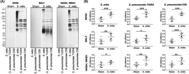
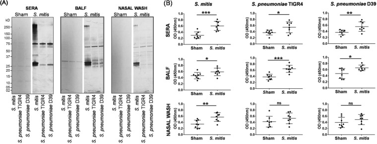
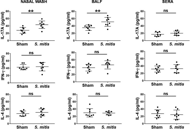
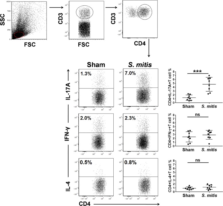
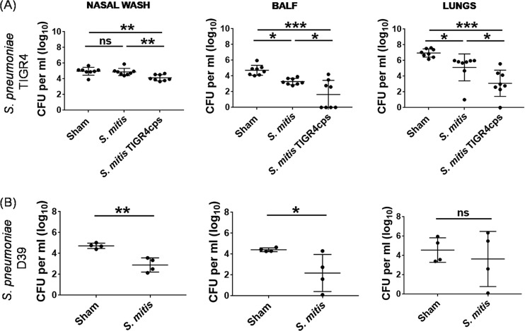
Streptococcus pneumoniae is a bacterial pathogen that causes various diseases of public health concern worldwide. Current pneumococcal vaccines target the capsular polysaccharide surrounding the cells. However, only up to 13 of more than 90 pneumococcal capsular serotypes are represented in the current conjugate vaccines. In this study, we used two experimental approaches to evaluate the potential of Streptococcus mitis, a commensal that exhibits immune cross-reactivity with S. pneumoniae, to confer protective immunity to S. pneumoniae lung infection in mice. First, we assessed the immune response and protective effect of wild-type S. mitis against lung infection by S. pneumoniae strains D39 (serotype 2) and TIGR4 (serotype 4). Second, we examined the ability of an S. mitis mutant expressing the S. pneumoniae type 4 capsule (S. mitis TIGR4cps) to elicit focused protection against S. pneumoniae TIGR4. Our results showed that intranasal immunization of mice with S. mitis produced significantly higher levels of serum IgG and IgA antibodies reactive to both S. mitis and S. pneumoniae, as well as enhanced production of interleukin 17A (IL-17A), but not gamma interferon (IFN-γ) and IL-4, compared with control mice. The immunization resulted in a reduced bacterial load in respiratory tissues following lung infection with S. pneumoniae TIGR4 or D39 compared with control mice. With S. mitis TIGR4cps, protection upon challenge with S. pneumoniae TIGR4 was superior. Thus, these findings show the potential of S. mitis to elicit natural serotype-independent protection against two pneumococcal serotypes and to provide the benefits of the well-recognized protective effect of capsule-targeting vaccines.IMPORTANCEStreptococcus pneumoniae causes various diseases worldwide. Current pneumococcal vaccines protect against a limited number of more than 90 pneumococcal serotypes, accentuating the urgent need to develop novel prophylactic strategies. S. pneumoniae and the commensal Streptococcus mitis share immunogenic characteristics that make S. mitis an attractive vaccine candidate against S. pneumoniae In this study, we evaluated the potential of S. mitis and its mutant expressing pneumococcal capsule type 4 (S. mitis TIGR4cps) to induce protection against S. pneumoniae lung infection in mice. Our findings show that intranasal vaccination with S. mitis protects against S. pneumoniae strains D39 (serotype 2) and TIGR4 (serotype 4) in a serotype-independent fashion, which is associated with enhanced antibody and T cell responses. Furthermore, S. mitis TIGR4cps conferred additional protection against S. pneumoniae TIGR4, but not against D39. The findings highlight the potential of S. mitis to generate protection that combines both serotype-independent and serotype-specific responses.
Keywords: Streptococcus mitis; Streptococcus pneumoniae; commensals; infection; pneumonia; vaccines.
Copyright © 2019 American Society for Microbiology.
Figures

Similar articles
-
Vaccination With the Commensal Streptococcus mitis Expressing Pneumococcal Serotype 5 Capsule Elicits IgG/IgA and Th17 Responses Against Streptococcus pneumoniae.Front Immunol. 2021 Apr 19;12:676488. doi: 10.3389/fimmu.2021.676488. eCollection 2021. Front Immunol. 2021. PMID: 33953733 Free PMC article.
-
Mucosal and systemic immunization with a novel attenuated pneumococcal vaccine candidate confer serotype independent protection against Streptococcus pneumoniae in mice.Vaccine. 2014 Jul 16;32(33):4179-88. doi: 10.1016/j.vaccine.2014.05.019. Epub 2014 Jun 2. Vaccine. 2014. PMID: 24945468
-
Serotype-Independent Protection Against Invasive Pneumococcal Infections Conferred by Live Vaccine With lgt Deletion.Front Immunol. 2019 May 29;10:1212. doi: 10.3389/fimmu.2019.01212. eCollection 2019. Front Immunol. 2019. PMID: 31191555 Free PMC article.
-
Streptococcus pneumoniae serotype 19A: worldwide epidemiology.Expert Rev Vaccines. 2017 Oct;16(10):1007-1027. doi: 10.1080/14760584.2017.1362339. Epub 2017 Aug 28. Expert Rev Vaccines. 2017. PMID: 28783380 Review.
-
Understanding host immune responses to pneumococcal proteins in the upper respiratory tract to develop serotype-independent pneumococcal vaccines.Expert Rev Vaccines. 2020 Oct;19(10):959-972. doi: 10.1080/14760584.2020.1843433. Epub 2020 Nov 8. Expert Rev Vaccines. 2020. PMID: 33107359 Review.
Cited by
-
Is the oral microbiome a source to enhance mucosal immunity against infectious diseases?NPJ Vaccines. 2021 Jun 2;6(1):80. doi: 10.1038/s41541-021-00341-4. NPJ Vaccines. 2021. PMID: 34078913 Free PMC article. Review.
-
Human Serum Supplementation Promotes Streptococcus mitis Growth and Induces Specific Transcriptomic Responses.Microbiol Spectr. 2023 Jun 15;11(3):e0512922. doi: 10.1128/spectrum.05129-22. Epub 2023 Apr 4. Microbiol Spectr. 2023. PMID: 37014220 Free PMC article.
-
Episodic Aspiration with Oral Commensals Induces a MyD88-dependent, Pulmonary T-Helper Cell Type 17 Response that Mitigates Susceptibility to Streptococcus pneumoniae.Am J Respir Crit Care Med. 2021 May 1;203(9):1099-1111. doi: 10.1164/rccm.202005-1596OC. Am J Respir Crit Care Med. 2021. PMID: 33166473 Free PMC article.
-
Panel 4: Recent advances in understanding the natural history of the otitis media microbiome and its response to environmental pressures.Int J Pediatr Otorhinolaryngol. 2020 Mar;130 Suppl 1(Suppl 1):109836. doi: 10.1016/j.ijporl.2019.109836. Epub 2019 Dec 18. Int J Pediatr Otorhinolaryngol. 2020. PMID: 31879084 Free PMC article. Review.
-
Treatment of Mouse Infants with Amoxicillin, but Not the Human Milk-Derived Antimicrobial HAMLET, Impairs Lung Th17 Responses.Antibiotics (Basel). 2023 Feb 20;12(2):423. doi: 10.3390/antibiotics12020423. Antibiotics (Basel). 2023. PMID: 36830333 Free PMC article.
References
-
- O'Brien KL, Wolfson LJ, Watt JP, Henkle E, Deloria-Knoll M, McCall N, Lee E, Mulholland K, Levine OS, Cherian T, Hib and Pneumococcal Global Burden of Disease Study Team 2009. Burden of disease caused by Streptococcus pneumoniae in children younger than 5 years: global estimates. Lancet 374:893–902. doi:10.1016/S0140-6736(09)61204-6. - DOI - PubMed
-
- World Health Organization. 2017. WHO publishes list of bacteria for which new antibiotics are urgently needed. http://www.who.int/news-room/detail/27-02-2017-who-publishes-list-of-bac.... World Health Organization, Geneva, Switzerland.
Publication types
MeSH terms
Substances
LinkOut - more resources
Full Text Sources
Other Literature Sources
Medical
Miscellaneous
