. 2019 Jan 25;10(2):74.
doi: 10.1038/s41419-019-1328-4.

SETD3 is a positive regulator of DNA-damage-induced apoptosis

Affiliations

SETD3 is a positive regulator of DNA-damage-induced apoptosis

Elina Abaev-Schneiderman et al. Cell Death Dis. .

Abstract

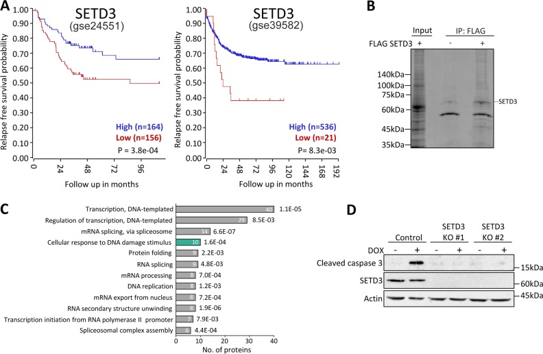
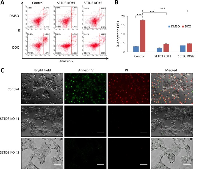
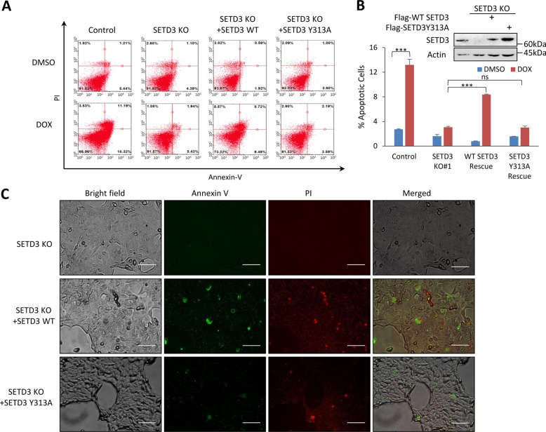
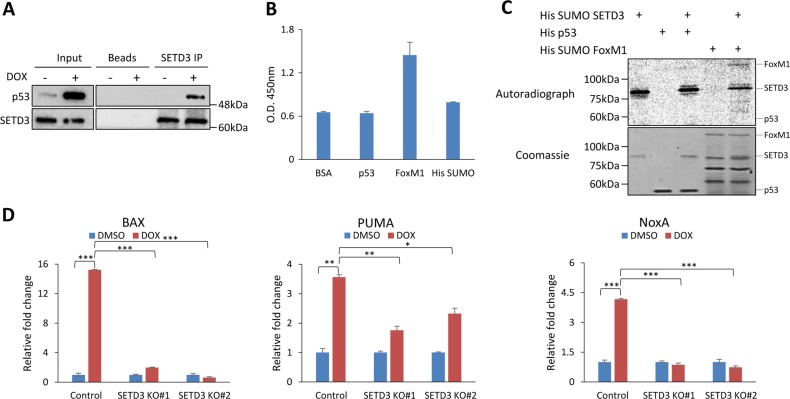
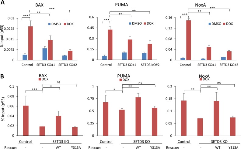
SETD3 is a member of the protein lysine methyltransferase (PKMT) family, which catalyzes the addition of methyl group to lysine residues. However, the protein network and the signaling pathways in which SETD3 is involved remain largely unexplored. In the current study, we show that SETD3 is a positive regulator of DNA-damage-induced apoptosis in colon cancer cells. Our data indicate that depletion of SETD3 from HCT-116 cells results in a significant inhibition of apoptosis after doxorubicin treatment. Our results imply that the positive regulation is sustained by methylation, though the substrate remains unknown. We present a functional cross-talk between SETD3 and the tumor suppressor p53. SETD3 binds p53 in cells in response to doxorubicin treatment and positively regulates p53 target genes activation under these conditions. Mechanistically, we provide evidence that the presence of SETD3 and its catalytic activity is required for the recruitment of p53 to its target genes. Finally, Kaplan-Meier survival analysis, of two-independent cohorts of colon cancer patients, revealed that low expression of SETD3 is a reliable predictor of poor survival in these patients, which correlates with our findings. Together, our data uncover a new role of the PKMT SETD3 in the regulation of p53-dependent activation of apoptosis in response to DNA damage.


Conflict of interest statement

The authors declare that they have no conflict of interest.

Figures

Fig. 1
Fig. 1. SETD3 interacts with DNA-damage associated proteins.
a Kaplan–Meier survival recurrence-free survival of 333 (gse24551) (Left) and of 566 (gse39582) (Right) colorectal cancer patients. High SETD3 expression represented in the blue line, while low expression of SETD3 is in red. b Coomassie staining of HCT-116 cells lysate ± overexpression of FLAG-SETD3 followed by immunoprecipitation with FLAG antibody conjugated beads. Input lane is of FLAG-SETD3 overexpressed cell lysate. c GO biological processes analysis of proteins identified in the mass spectrometry experiment. d HCT-116 control and SETD3 KO cells treated with DOX, followed by western blot with the indicated antibodies
Fig. 2
Fig. 2. SETD3 positively regulates DOX-induced apoptosis.
a FACS analysis of KO and control cells post DOX or vehicle treatment. b Quantification of apoptotic cells percentage of three-independent FACS analyses, ***p ≤ 0.001. c Control and KO cells were stained with FITC-Annexin V and PI post treatment with DOX and viewed under fluorescent microscope (scale bar signifies 100 µM, pictures were taken under ×20 magnification)
Fig. 3
Fig. 3. DNA-damage-induced apoptosis is SETD3 and methylation dependent.
a FACS analysis of control, KO, WT SETD3 rescue and catalytic inactive (Y313A) SETD3 rescue cells, post DOX or vehicle treatment. b Quantification of apoptotic cells percentage of three-independent FACS analyses, ***p ≤ 0.001, ns p > 0.05. Western blot of the control, KO and rescued cells lysate. c SETD3 KO and rescued (with WT or Y313A) cells were stained with FITC-Annexin V and PI post treatment with DOX and viewed under fluorescent microscope (scale bar signifies 100 µM, pictures were taken under ×20 magnification)
Fig. 4
Fig. 4. SETD3-positively regulates the activation of DNA-damage-induced apoptosis target genes.
a Immunoprecipitation of endogenous SETD3 from whole-cell lysate (HCT-116 cells) followed by western blot with the indicated antibodies. b ELISA experiment between His-p53 and His-SUMO SETD3, His-SUMO FoxM1 used as positive control. c In vitro methylation assay between His-SUMO SETD3 and His-p53. Using His-SUMO FoxM1 as positive control. d qPCR analysis of extracted mRNA from HCT-116 control and KO cells, as indicated, post DOX or vehicle treatment. Graphs show the relative expression levels (normalized to GAPDH) of the indicated genes. *p ≤ 0.05, **p ≤ 0.01, ***p ≤ 0.001
Fig. 5
Fig. 5. SETD3 catalytic activity is required for p53 recruitment to its target genes following DOX treatment.
a Chromatin immunoprecipitation (ChIP) assay of control and SETD3 KO HCT-116 cells treated with DOX or vehicle. DNA fragments were immunoprecipitated with p53 antibody. Values were compared to input samples. *p ≤ 0.05, **p ≤ 0.01, ***p ≤ 0.001. b Chromatin immunoprecipitation (ChIP) assay of control, SETD3 KO and rescued (with WT or Y313A) cells, post DOX treatment. DNA fragments were immunoprecipitated with p53 antibody. Values were compared to input samples. *p ≤ 0.05, **p ≤ 0.01, ***p ≤ 0.001

References

    1. Thompson CB. Apoptosis in the pathogenesis and treatment of disease. Science. 1995;267:1456–1462. doi: 10.1126/science.7878464. - DOI - PubMed
    1. Coutts AS, La Thangue NB. The p53 response: emerging levels of co-factor complexity. Biochem. Biophys. Res. Commun. 2005;331:778–785. doi: 10.1016/j.bbrc.2005.03.150. - DOI - PubMed
    1. Thornborrow EC, Manfredi JJ. The tumor suppressor protein p53 requires a cofactor to activate transcriptionally the human BAX promoter. J. Biol. Chem. 2001;276:15598–15608. doi: 10.1074/jbc.M011643200. - DOI - PubMed
    1. Nakano K, Vousden KH. PUMA, a novel proapoptotic gene, is induced by p53. Mol. Cell. 2001;7:683–694. doi: 10.1016/S1097-2765(01)00214-3. - DOI - PubMed
    1. Shibue T, et al. Integral role of Noxa in p53-mediated apoptotic response. Genes Dev. 2003;17:2233–2238. doi: 10.1101/gad.1103603. - DOI - PMC - PubMed

Publication types

MeSH terms