. 2019 Mar;15(1):27-35.
doi: 10.1007/s11302-019-09646-5. Epub 2019 Jan 25.

Evidence for detection of rat P2X4 receptor expressed on cells by generating monoclonal antibodies recognizing the native structure

Affiliations

Evidence for detection of rat P2X4 receptor expressed on cells by generating monoclonal antibodies recognizing the native structure

Tatsuhiro Igawa et al. Purinergic Signal. 2019 Mar.

Abstract

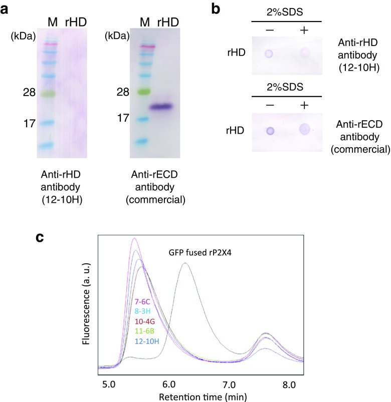
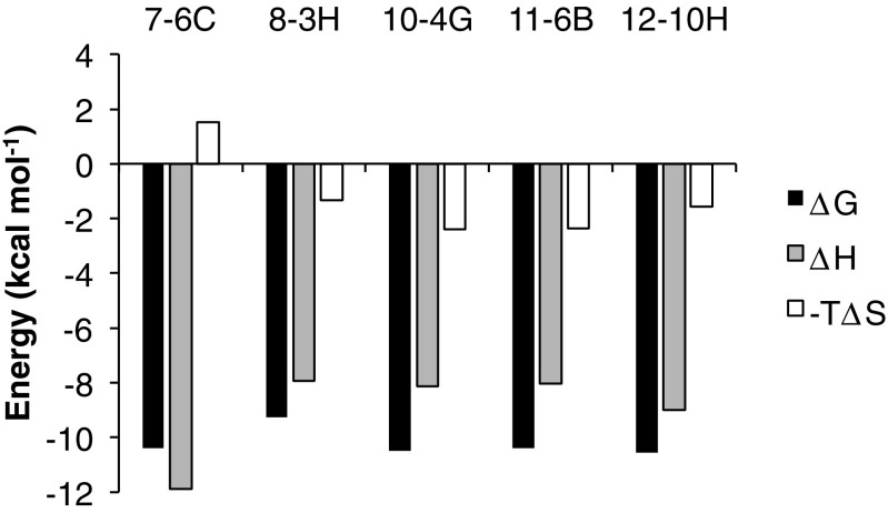
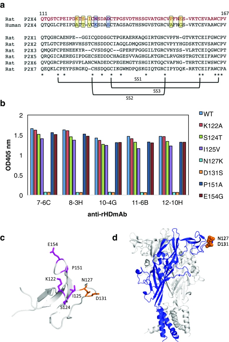
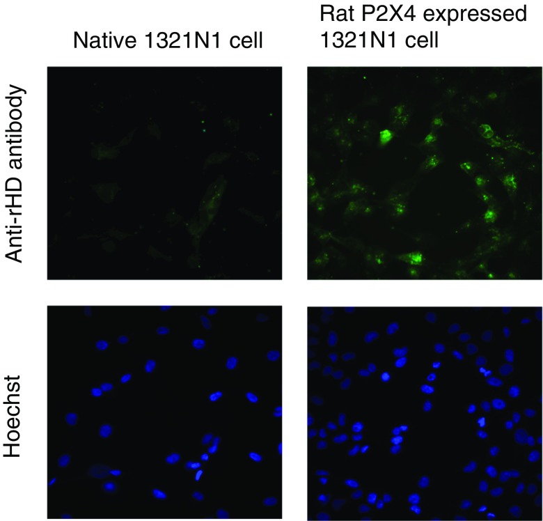
P2X purinergic receptors are ATP-driven ionic channels expressed as trimers and showing various functions. A subtype, the P2X4 receptor present on microglial cells is highly involved in neuropathic pain. In this study, in order to prepare antibodies recognizing the native structure of rat P2X4 (rP2X4) receptor, we immunized mice with rP2X4's head domain (rHD, Gln111-Val167), which possesses an intact structure stabilized by S-S bond formation (Igawa and Abe et al. FEBS Lett. 2015), as an antigen. We generated five monoclonal antibodies with the ability to recognize the native structure of its head domain, stabilized by S-S bond formation. Site-directed mutagenesis revealed that Asn127 and Asp131 of the rHD, in which combination of these amino acid residues is only conserved in P2X4 receptor among P2X family, were closely involved in the interaction between rHD and these antibodies. We also demonstrated the antibodies obtained here could detect rP2X4 receptor expressed in 1321N1 human astrocytoma cells.

Keywords: FSEC; Monoclonal antibody; Neuropathic pain; P2X4 receptor.


Conflict of interest statement

Conflicts of interest

Tatsuhiro Igawa declares that he has no conflict of interest.

Shuhei Kishikawa declares that he has no conflict of interest.

Yoshito Abe declares that he has no conflict of interest.

Tomohiro Yamashita declares that he has no conflict of interest.

Saki Nagai declares that she has no conflict of interest.

Mitsunori Shiroishi declares that he has no conflict of interest.

Chinatsu Shinozaki declares that he has no conflict of interest.

Hiroyuki Tanaka declares that he has no conflict of interest.

Hidetoshi Tozaki-Saitoh declares that he has no conflict of interest.

Makoto Tsuda declares that he has no conflict of interest.

Kazuhide Inoue declares that he has no conflict of interest.

Tadashi Ueda declares that he has no conflict of interest.

Ethical approval

All animal experiments were conducted according to the relevant national and international guidelines in the Act on Welfare and Management of Animals (Ministry of Environment of Japan) and the Regulation of Laboratory Animals (Kyushu University), and under the protocols approved by the Institutional Animal Care and Use Committee review panels at Kyushu University.

Figures

Fig. 1
Fig. 1
Screening of monoclonal antibodies. a Western blotting for rHD using anti-rHD antibody 12-10H (left) and anti-rECD antibody (right). b Dot blot of rHD in the presence (right) or absence (left) of 2% SDS using anti-rHD antibody 12-10H (upper) and anti-rECD antibody (lower). c GFP-fused rat P2X4 in the absence (black) or the presence of 7-6C (magenta), 8-3H (cyan), 10-4G (brown), 11-6B (green), and 12-10H (indigo) was monitored by fluorescence-detection size-exclusion chromatography (FSEC). FSEC was performed with the Superdex 200 5/150 GL at flow rate 0.5 ml/min. The fluorescence was detected at 525 nm with excitation at 490 nm
Fig. 2
Fig. 2
Thermodynamics of binding of each monoclonal antibody to rHD at 25 °C. ΔG (black bar), ΔH (dotted bar), and −TΔS (white bar) are shown
Fig. 3
Fig. 3
Epitope of rP2X4 for anti-rHD antibody. a Sequence alignment of the head domain of human P2X4 and rat P2X1~P2X7. The mutation sites were enclosed in squares. Conserved amino acids are indicated by asterisks and three S-S bond formations are represented. b The results of ELISA for rHD mutants (K122A, S124T, I125V, N127K, D131S, P151A, and E154G) using each monoclonal antibody as a primary antibody and alkaline phosphatase–conjugated anti-mouse IgG as a secondary antibody. c Mutation sites (stick model) were mapped on the structure of rHD. The epitope is shown in blue. d The epitope is indicated on the homology model of the rP2X4 trimer. One monomer in the rP2X4 trimer is shown in blue
Fig. 4
Fig. 4
Disulfide bond contribution of anti-rHD antibody binding to rHD. a Disulfide bonds of rHD are depicted on the structure of rHD. Cysteines are shown in yellow. b Protease sensitivity of S-S bond deletion mutants of rHD, ΔSS1 (C116A/C165A), ΔSS2 (C126A/C149A), and ΔSS3 (C132A/C159A), and wild-type rHD. Proteins (25 μM) were treated with 100 μg/ml cathepsin B for the indicated times at 25 °C. Under reducing conditions, the sample was incubated with 52 mmol/l 2-mercaptoethanol for 40 °C and 90 min before digestion. The presence of protein in the gels was detected with silver staining. c The results of ELISA for S-S bond deletion rHD mutants, ΔSS1 (C116A/C165A), ΔSS2 (C126A/C149A), and ΔSS3 (C132A/C159A), using each monoclonal antibody
Fig. 5
Fig. 5
Detection of rP2X4 expressed on the cell by monoclonal antibody. Anti-rHD monoclonal antibody (12-10H, 10 μg/ml) and Alexa488-conjugated anti-mouse IgG were used for staining rP2X4 expressed on the 1321N1 cell (upper right). A native 1321N1 cell was also stained using both antibodies as a control (upper left). Hoechst was used to stain nuclei simultaneously (lower)

Similar articles

Cited by

References

    1. Watkins LR, Maier SF. Glia: a novel drug discovery target for clinical pain. Nat Rev Drug Discov. 2003;2(12):973–985. doi: 10.1038/nrd1251. - DOI - PubMed
    1. Watkins LR, Milligan ED, Maier SF. Spinal cord glia: new players in pain. Pain. 2001;93(3):201–205. doi: 10.1016/S0304-3959(01)00359-1. - DOI - PubMed
    1. Watkins LR, Milligan ED, Maier SF. Glial activation: a driving force for pathological pain. Trends Neurosci. 2001;24(8):450–455. doi: 10.1016/S0166-2236(00)01854-3. - DOI - PubMed
    1. Tsuda M, Shigemoto-Mogami Y, Koizumi S, Mizokoshi A, Kohsaka S, Salter MW, Inoue K. P2X4 receptors induced in spinal microglia gate tactile allodynia after nerve injury. Nature. 2003;424(6950):778–783. doi: 10.1038/nature01786. - DOI - PubMed
    1. Coull JA, Beggs S, Boudreau D, Boivin D, Tsuda M, Inoue K, Gravel C, Salter MW, De Koninck Y. BDNF from microglia causes the shift in neuronal anion gradient underlying neuropathic pain. Nature. 2005;438(7070):1017–1021. doi: 10.1038/nature04223. - DOI - PubMed

Publication types

Substances

LinkOut - more resources