Review
. 2019 Nov;128(3):288-297.
doi: 10.1016/j.ymgme.2018.12.008. Epub 2018 Dec 27.

Congenital erythropoietic porphyria: Recent advances

Affiliations
Review

Congenital erythropoietic porphyria: Recent advances

Angelika L Erwin et al. Mol Genet Metab. 2019 Nov.

Abstract

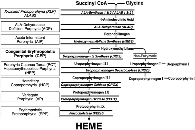
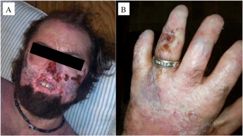
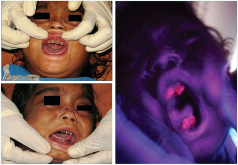
Congenital erythropoietic porphyria (CEP) is a rare autosomal recessive disorder characterized by photosensitivity and by hematologic abnormalities in affected individuals. CEP is caused by mutations in the uroporphyrinogen synthase (UROS) gene. In three reported cases, CEP has been associated with a specific X-linked GATA1 mutation. Disease-causing mutations in either gene result in absent or markedly reduced UROS enzymatic activity. This in turn leads to the accumulation of the non-physiologic and photoreactive porphyrinogens, uroporphyrinogen I and coproporphyrinogen I, which damage erythrocytes and elicit a phototoxic reaction upon light exposure. The clinical spectrum of CEP depends on the level of residual UROS activity, which is determined by the underlying pathogenic loss-of-function UROS mutations. Disease severity ranges from non-immune hydrops fetalis in utero to late-onset disease with only mild cutaneous involvement. The clinical characteristics of CEP include exquisite photosensitivity to visible light resulting in bullous vesicular lesions which, when infected lead to progressive photomutilation of sun-exposed areas such as the face and hands. In addition, patients have erythrodontia (brownish discoloration of teeth) and can develop corneal scarring. Chronic transfusion-dependent hemolytic anemia is common and leads to bone marrow hyperplasia, which further increases porphyrin production. Management of CEP consists of strict avoidance of exposure to visible light with sun-protective clothing, sunglasses, and car and home window filters. Adequate care of ruptured vesicles and use of topical antibiotics is indicated to prevent superinfections and osteolysis. In patients with symptomatic hemolytic anemia, frequent erythrocyte cell transfusions may be necessary to suppress hematopoiesis and decrease marrow production of the phototoxic porphyrins. In severe transfection-dependent cases, bone marrow or hematopoietic stem cell transplantation has been performed, which is curative. Therapeutic approaches including gene therapy, proteasome inhibition, and pharmacologic chaperones are under investigation.

Keywords: Cutaneous lesions, Heme biosynthetic pathway; Hemolysis; Phototoxicity; Porphyria.


Conflict of interest statement

Conflicts of Interest:

The authors report no relevant conflicts

Figures

Fig 1.
Fig 1.
The human biosynthetic pathway and the porphyrias resulting from the indicated heme biosynthetic enzyme defect. There are eight enzymatic steps in the conversion of glycine and succinyl-CoA to heme. The heme biosynthetic enzymes are italicized, their substrates and products are indicated, and the resulting porphyrias are shown in boxes. Note that there are two aminolevulinic acid synthase (ALAS) isozymes: a housekeeping enzyme, ALAS1, encoded by a gene that is regulated by negative feedback repression by heme, and an erythroid specific enzyme, ALAS2, that is regulated by the iron response proteins and erythroid transcription binding proteins.
Fig 2.
Fig 2.
Uroporphyrinogen synthase (UROS) normally converts hydroxymethylbilane (HMB) to uroporphyrinogen III. When the UROS activity is markedly decreased, HMB is non-enzymatically converted to uroporphyrinogen I, which is then enzymatically converted to oproporphyrinogen I by uroporphyrinogen decarboxylase. The accumulated uroporphyrinogen I and coproporphyrinogen I are oxidized to their respective porphyrins, which are photoactive and cause the sun/light-induced hemolysis and cutaneous manifestations of CEP.
Fig 3.
Fig 3.
Diaper of an infant with CEP. The patient’s urine has stained the diaper a reddish color due to the accumulated uroporphyrin I and coproporphyrin I (left). The accumulated porphyrins fluoresce when illuminated with ultraviolet light (right) [95].
Fig 4.
Fig 4.
Organization of the UROS gene. The UROS gene has unique housekeeping (PH) and erythroid-specific (PE) promoters. The dotted lines indicate the exons transcribed by each promoter. However, both promoters encode the same enzyme polypeptide.
Fig 5.
Fig 5.
Molecular lesions in the UROS gene causing CEP. Coding exons are shown as solid black rectangles. ATG is the initiation of translation codon of the housekeeping and erythroid transcripts. Missense and nonsense mutations are indicated by the one-letter amino acid code and codon position, e.g. Q155X = glutamine codon (Q) at position 155 replaced by a termination codon (X). Deletions are indicated by Δ and insertions by ins. Note that the mutation encoding C73R is the most common lesion found in ~30–35% of reported patients [10].
Fig 6.
Fig 6.
Partial sequence of the human and murine UROS erythroid-specific promoters showing the GATA1 and CP2 transcription factor binding sites. The location and orientation (<, >) of the GATA1, E-box, and CP2 erythroid binding elements are indicated, as are the four novel promoter mutations causing CEP (−70C, −76A, −86A and −90A). Dots are placed every tenth nucleotide. The homologous CP2 site from the α-globin gene promoter and the GATA1 site from the human stem cell leukemia (SCL) gene promoter are shown.
Fig 7.
Fig 7.
Male 34 year-old CEP patient with severe cutaneous manifestations of face (left) and hand (right). Note the extensive skin erosions in the face, destruction of the nasal cartilage, sclerodermoid changes of the mouth, and erythrodontia. The patient also has significant shortening of the digits due to photomutilation and contractures of the joints, along with erosions and scleroderma-like thickening of the skin.
Fig 8.
Fig 8.
Photographs showing the brownish color of the teeth on an infant with CEP in daylight (left) and fluorescing when illuminated with ultraviolet light from a Woods lamp (right) [96].

References

    1. Anderson KE, Sassa S, Bishop DF, Desnick RJ, Disorders of heme biosynthesis: X-linked sideroblastic anemia and the porphyrias In: Metabolic The and Molecular Bases of Inherited Disease, 8th (ed), Scriver CR, Beaudet AL, Sly WS and Valle D, (eds), New York (NY), McGraw-Hill, (2014) 2961–3062. http://ommbid.mhmedical.com/content.aspx?bookid=971&Sectionid=62638866.
    1. Puy H, Gouya L, Deybach JC, Porphyrias. Lancet 375 (2010) 924–937. - PubMed
    1. Balwani M, Desnick RJ, The porphyrias: advances in diagnosis and treatment. Blood 120 (2012) 4496–4504. - PMC - PubMed
    1. Romeo G, Levin EY, Uroporphyrinogen 3 cosynthetase in human congenital erythropoietic porphyria. Proc Natl Acad Sci U S A 63 (1969) 856–863. - PMC - PubMed
    1. Bickers DR, Frank J, The Porphyrias. In: Fitzpatrick’s Dermatology in General Medicine, Goldsmith LA, Katz SI, Gilchrest BA, Paller AS, Leffell DJ, Wolff K (eds), 8th (ed), McGraw-Hill, New York (NY), Chapter 132, (2012) 1679 https://accessmedicine.mhmedical.com/Content.aspx?bookId=392&sectionId=4....

Publication types