. 2019 Apr 1:189:445-458.
doi: 10.1016/j.neuroimage.2019.01.059. Epub 2019 Jan 24.

Magnetoencephalography and the infant brain

Affiliations

Magnetoencephalography and the infant brain

Yu-Han Chen et al. Neuroimage. .

Abstract

Magnetoencephalography (MEG) is a non-invasive neuroimaging technique that provides whole-head measures of neural activity with millisecond temporal resolution. Over the last three decades, MEG has been used for assessing brain activity, most commonly in adults. MEG has been used less often to examine neural function during early development, in large part due to the fact that infant whole-head MEG systems have only recently been developed. In this review, an overview of infant MEG studies is provided, focusing on the period from birth to three years. The advantages of MEG for measuring neural activity in infants are highlighted (See Box 1), including the ability to assess activity in brain (source) space rather than sensor space, thus allowing direct assessment of neural generator activity. Recent advances in MEG hardware and source analysis are also discussed. As the review indicates, efforts in this area demonstrate that MEG is a promising technology for studying the infant brain. As a noninvasive technology, with emerging hardware providing the necessary sensitivity, an expected deliverable is the capability for longitudinal infant MEG studies evaluating the developmental trajectory (maturation) of neural activity. It is expected that departures from neuro-typical trajectories will offer early detection and prognosis insights in infants and toddlers at-risk for neurodevelopmental disorders, thus paving the way for early targeted interventions.

Keywords: Auditory; Development; Infant; MEG; Somatosensory; Visual.


Figures

Fig. 1.
Fig. 1.
Photos of existing pediatric MEG systems. (A) SARA system (VSM Med Tech Ltd.) being used for fetal MEG recording. (B) SARA system with cradle attachment adapted for infant recording (both images from Holst et al., 2005). (C) BabySQUID system with partial head coverage (Tristan Technologies Inc.) (D) KIT 151-channel whole-head system (Model PQ1151R; Yokogawa/KIT; image adapted from Kikuchi et al., 2013). (E) Artemis 123 whole-head system (Tristan Technologies Inc.). (F) Baby-MEG 375-channel whole-head system (Tristan Technologies Inc.).
Fig. 2.
Fig. 2.
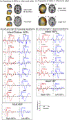
Examples of (a) Topoplots of auditory evoked fields (AEFs) from a representative 5-month-old infant and from an adult, (b) Source waveforms from left and right STG (c) Topoplots of visual evoked fiExamples of (a) Topoplots of auditory evoked fields (AEFs) from a representative 5-month-old infant and from an adult, (b) Source waveforms from left and right STG (c) Topoplots of visual evoked temis 123 whole-head system (Tristan rphological analogies can be seen in the infant, young children, and adult waveforms, although for auditory responses the component latencies (marked with dash lines) have strikingly longer latencies in infants. For example, the “infant M50” occurs at 197 ms for a 2-month-old infant, and the M50 latencies decrease to 108 ms in a representative 6-year-old child, and 66 ms in a representative adult. By contrast VEF latencies appear to mature faster toward the adult values compared to AEF.
Fig. 3.
Fig. 3.
Scatterplot of auditory M50 latencies from 6-month-old to 65-year-old subjects. The analogies between infant and child and adult morphologies in auditory evoked fields permit assessment of the lifespan trajectory of component latencies. This response is robustly observed across different hardware platforms with findings suggesting a continuous shortening of “M50” latency from infancy to adulthood. The absence of discontinuity indicates the potential to track a component latency across the lifespan despite differing hardware, offering potential for multicenter implementations.
Fig. 4.
Fig. 4.
An example of (a) Face versus non-face contrast of whole-brain activity in five 6-month-old infants, and (b) source waveforms from left and right fusiform gyrus (FFG) to face and non-face stimuli. Greater activity (indicated in red) was observed in response to face versus non-face stimuli in FFG (200–400 ms post-stimulus), V1, temporal pole and frontal areas. FFG source waveforms showed that right FFG activity in response to face stimuli peaked at ~300 ms and ~400 ms, and left FFG activity to face stimuli peaked at ~350 ms.

References

    1. Akiyama LF, Richards TR, Imada T, Dager SR, Wroblewski L, Kuhl PK, 2013. Age-specific average head template for typically developing 6-month-old infants. PLoS One 8 e73821. - PMC - PubMed
    1. Anderson AL, Thomason ME, 2013. Functional plasticity before the cradle: a review of neural functional imaging in the human fetus. Neurosci. Biobehav. Rev 37, 2220–2232. - PubMed
    1. Association AP, 2000. Diagnostic and Statistical Manual of Mental Disorders, 4 ed. American Psychiatric Association, Washington, DC.
    1. Bakhireva LN, Lowe JR, Gutierrez HL, Stephen JM, 2015. Ethanol, neurodevelopment, infant and child Health (ENRICH) prospective cohort: study design considerations. Adv Pediatr Res 2. - PMC - PubMed
    1. Barnet AB, 1975. Auditory evoked potentials during sleep in normal children from ten days to three years of age. Electroencephalogr. Clin. Neurophysiol 39, 29–41. - PubMed

Publication types

MeSH terms