. 2019 Feb 22:12:168-181.
doi: 10.1016/j.isci.2019.01.012. Epub 2019 Jan 11.

Imidazopyridines as Potent KDM5 Demethylase Inhibitors Promoting Reprogramming Efficiency of Human iPSCs

Affiliations

Imidazopyridines as Potent KDM5 Demethylase Inhibitors Promoting Reprogramming Efficiency of Human iPSCs

Yasamin Dabiri et al. iScience. .

Abstract

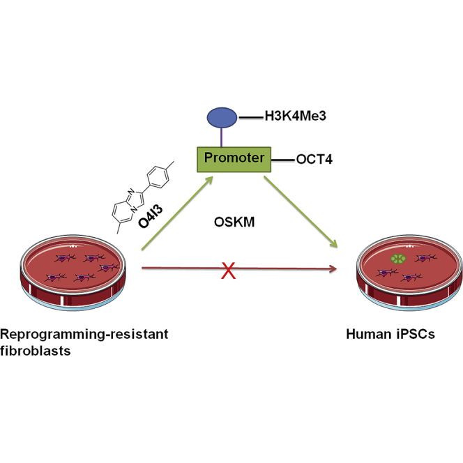
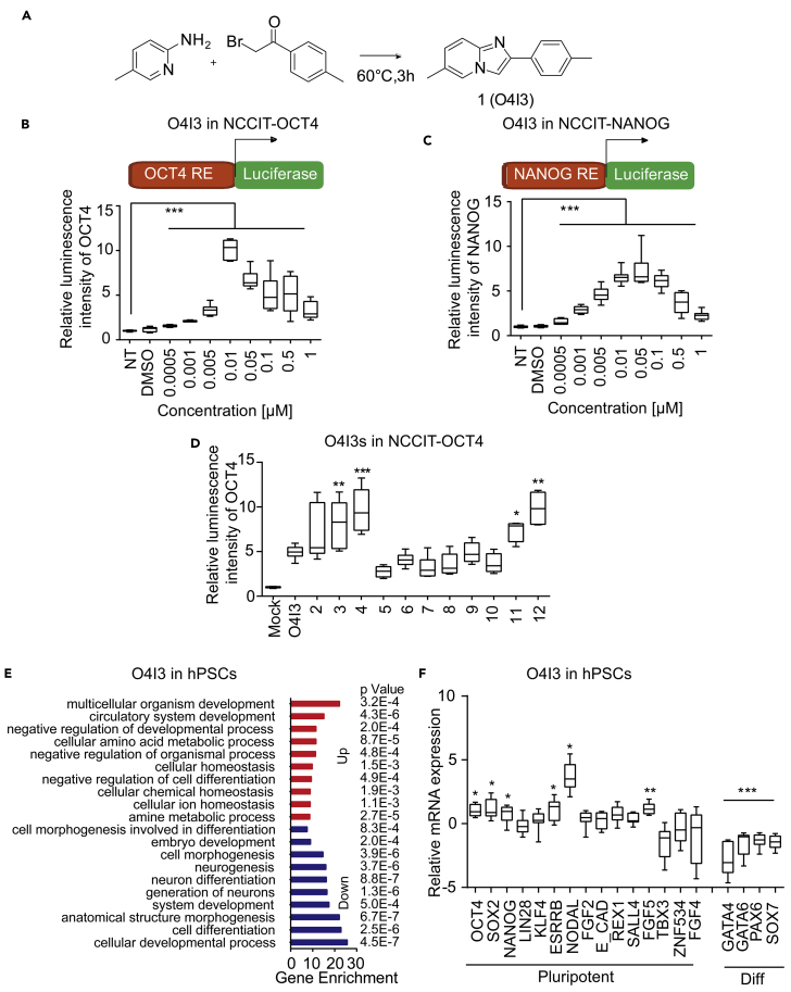
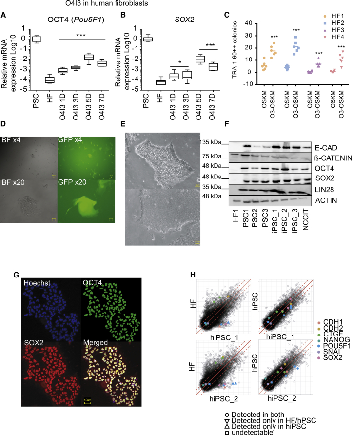
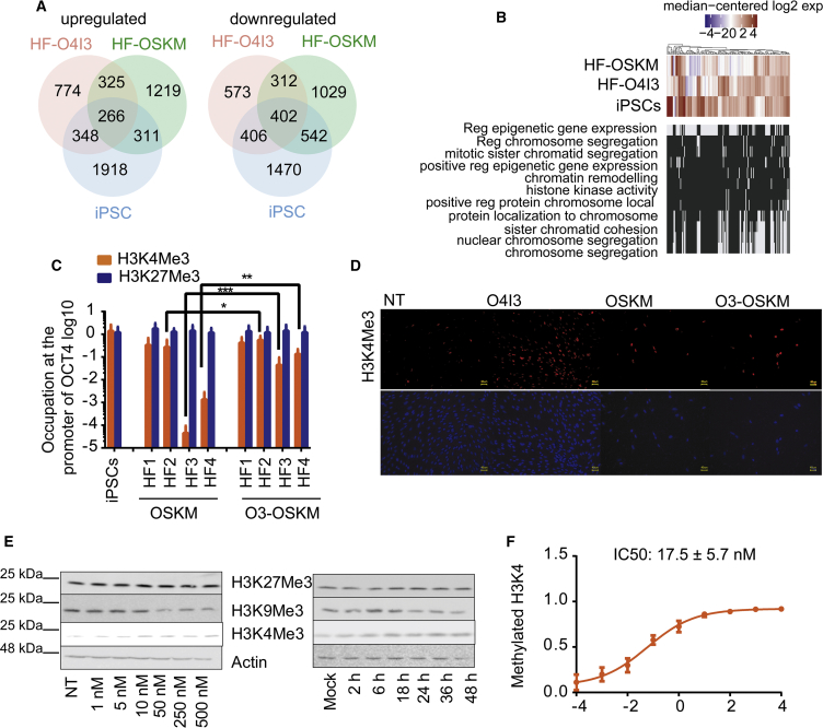
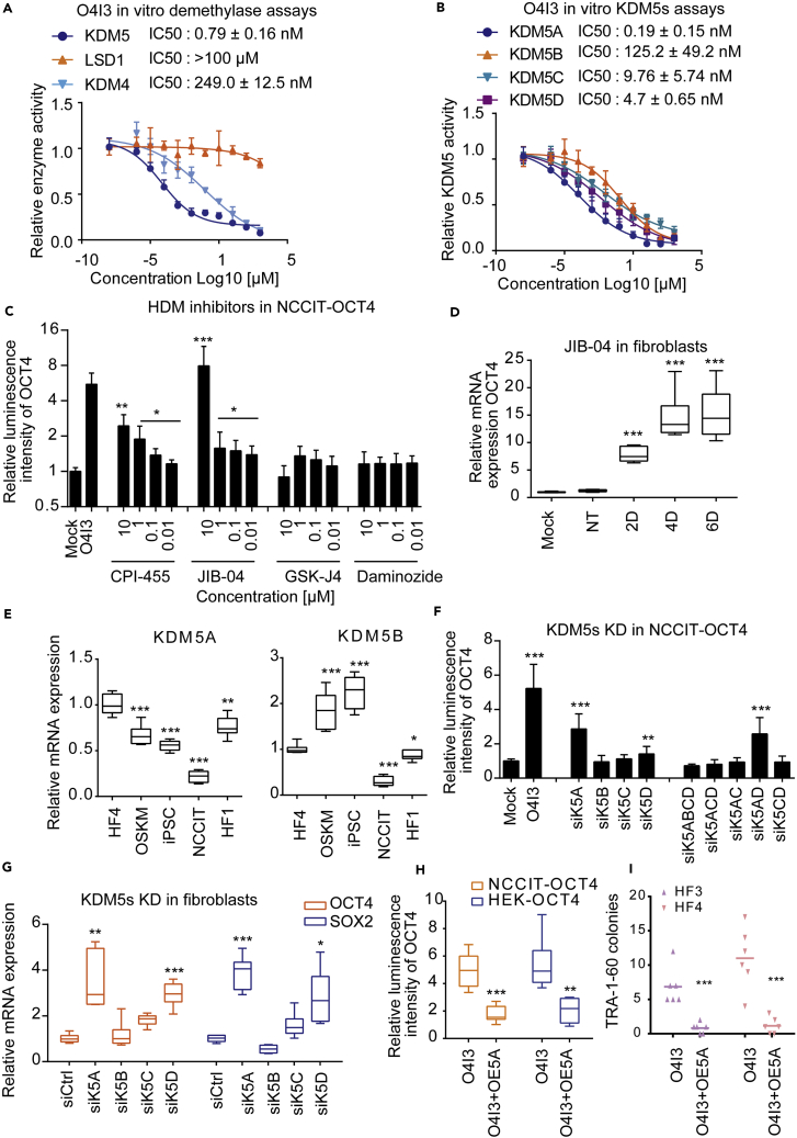
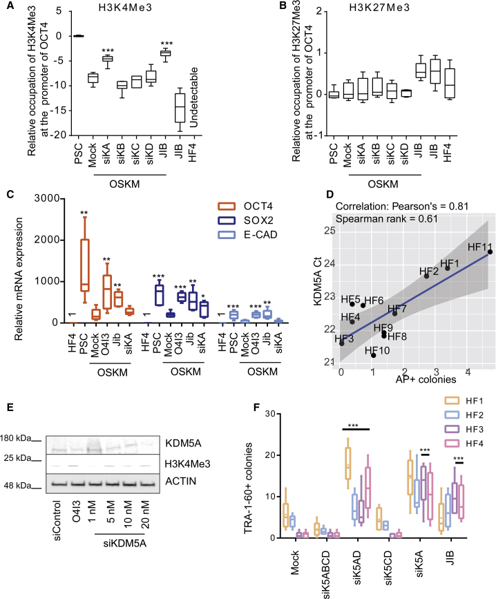
Pioneering human induced pluripotent stem cell (iPSC)-based pre-clinical studies have raised safety concerns and pinpointed the need for safer and more efficient approaches to generate and maintain patient-specific iPSCs. One approach is searching for compounds that influence pluripotent stem cell reprogramming using functional screens of known drugs. Our high-throughput screening of drug-like hits showed that imidazopyridines-analogs of zolpidem, a sedative-hypnotic drug-are able to improve reprogramming efficiency and facilitate reprogramming of resistant human primary fibroblasts. The lead compound (O4I3) showed a remarkable OCT4 induction, which at least in part is due to the inhibition of H3K4 demethylase (KDM5, also known as JARID1). Experiments demonstrated that KDM5A, but not its homolog KDM5B, serves as a reprogramming barrier by interfering with the enrichment of H3K4Me3 at the OCT4 promoter. Thus our results introduce a new class of KDM5 chemical inhibitors and provide further insight into the pluripotency-related properties of KDM5 family members.

Keywords: Biochemistry; Biological Sciences; Molecular Biology.


Figures

None
Graphical abstract
Figure 1
Figure 1
Identification of Imidazopyridine Analogs as OCT4-Inducing Compounds (A) Synthesis of the compound 1. (B and C) (B) O4I3 activates OCT4 in NCCIT-OCT4 response element (RE)-driven luciferase reporter cells and (C) NANOG in NCCIT-NANOG RE-driven luciferase reporter cells after 48h treatment. (D) Activity of O4I3 derivatives (50 nM) in NCCIT-OCT4 cells. (E) Functional enrichment analysis shows statistically significant enrichment in Gene Ontology terms related to pluripotency, development, and homeostasis in O4I3-treated PSCs for 24 h. Red, upregulated genes; blue, downregulated genes. p values represent values after multitest adjustment (see details in Transparent Methods). (F) qRT-PCR analysis of pluripotency- and differentiation (diff)-related gene expression in O4I3-treated hPSCs at a concentration of 10 nM for 48 h. In (B), (C), and (F) statistical significance was compared with mock (01% DMSO) treatment, whereas in (D) it was compared with O4I3 treatment using two-way ANOVA and a post-hoc Tukey test. Data are represented as mean ± SD. ***p<0.001, **p<0.01, *p<0.05.
Figure 2
Figure 2
O4I3 Improves OSKM-Induced Reprogramming (A and B) (A) O4I3 induces OCT4 and (B) SOX2 expression in human fibroblasts treated with O4I3 (50 nM) for the indicated time points (D, days). (C) Efficiency of OSKM-induced reprogramming in the presence or absence of O4I3 in human fibroblasts 1–4 (HF1–HF4). ESC-like colonies were detected by TRA-1-60 staining. (D) Reprogramming of resistant fibroblasts carrying LTR7-EGFP promoter. ESC-like GFP+ colonies were observed in bright field (BF) and GFP channel (GFP). (E) ESC-like colonies detected from newly generated hiPSC lines. (F) Expression of pluripotency-associated markers in the newly generated iPSC1, 2, and 3 when compared with hPSC1–3 as positive controls, determined by immunoblotting. (G) Co-expression of OCT4 and SOX2 in the newly generated iPSCs. (H) Scatterplots of gene expression data show high similarity between the newly generated iPSCs (hiPSC_1 and hiPSC_2) and hPSC (positive control), but not human fibroblasts (HF). All scatterplots are based on log2-transformed gene expression values. Red dashed lines depict the 2-fold change in expression values. In (A) and (B) statistical significance was compared with mock (0.1% DMSO) treatment in fibroblasts, whereas in (C) it was compared with the efficiency obtained by ectopic expression of OSKM using two-way ANOVA followed by a post-hoc Tukey test. Data are represented as mean ± SD. ***p<0.001, **p<0.01, *p<0.05.
Figure 3
Figure 3
O4I3 Is an Epigenetic Modulator (A) Venn diagram of up- and downregulated genes in O4I3 (50 nM)-treated human fibroblasts (HF) with ectopic expression of OCT4, SOX2, KLF4, and MYC (OSKM) as well as hiPSCs. HF-O4I3, HF cells treated with O4I3 (50 nM for 24 h); HF-OSKM, HF cells transfected with episomal OSKM; iPSCs, the newly generated iPSCs. Differential expression was calculated with respect to HF cells. (B) Comparison of expression profiles of genes involved in epigenetic modification, chromatin remodeling, and related biological processes. (C) Occupation of H3K4Me3 or H3K27Me3 at the promoter of OCT4 in HF cells transfected with OSKM alone or in the presence of 50 nM of O4I3 (O3-OSKM) for 3 days. (D) O4I3 elevates the global levels of H3K4Me3 detected by immunocytochemistry. Cells were treated with 250 nM O4I3 for 3 days. Scale bar, 40 μm. (E) Immunoblotting results show that O4I3 promotes global H3K4Me3 expression, whereas it represses H3K9Me3 protein levels in a concentration- and time-dependent manner. Cells were treated either with increasing concentrations of O4I3 for 24 h or with 250 nM of O4I3 for the indicated time points. (F) O4I3 protects the methylation of H3K4, as observed by an in vitro methylation assay using total nuclear extraction. In (C) statistical significance was compared with OSKM-treated fibroblasts using two-way ANOVA followed by a post-hoc Tukey test. Data are represented as mean ± SD. ***p<0.001, **p<0.01, *p<0.05.
Figure 4
Figure 4
O4I3 Is a Selective KDM5A Inhibitor (A) Comparison of O4I3 inhibitory effect on KDM5, LSD1, and KDM4 in vitro using the whole-cell nuclear extraction. (B) The inhibitory effect of O4I3 on the members of KDM5 family of demethylases isolated from cells. (C) A selective KDM5A inhibitor JIB-04 induces OCT4 expression in NCCIT-OCT4 cells. Four histone demethylase inhibitors (HDMs), namely, CPI-455, JIB-04, GSK-J4, and daminozide, were incubated with NCCIT-OCT4 reporter cells for 48 h. (D) JIB-04 (5 μM) induces OCT4 expression in fibroblasts at the indicated time points (D, days). (E) Comparison of KDM5A and KDM5B expression levels in fibroblast (HF1), resistant fibroblast (HF4), HF4 transfected with OSKM, iPSCs, and NCCIT. (F) Knockdown of KDM5A (si5A) activates OCT4 in NCCIT-OCT4 reporter cells. Cells were transfected with various KDM5 siRNA concentrations for 48 h. (G) Transient knockdown (48 h) of KDM5A induces OCT4 and SOX2 expression in fibroblasts. (H) Overexpression of KDM5A (OE5A) compromises the effect of O4I3 (100 nM, 48 h) in NCCIT-OCT4 and HEK-OCT4 cells. (I) The number of TRA-1-60-positive colonies was reduced in reprogramming-resistant fibroblasts with overexpression of KDM5A. In (C), (D), and (F) statistical significance was compared with mock (0.1% DMSO) treatment, in (G) with non-targeting siRNA and in (E) with HF4, whereas in (H) and (I) it was compared with OSKM-treated fibroblasts using two-way ANOVA followed by a post-hoc Tukey test. Data are represented as mean ± SD. ***p<0.001, **p<0.01, *p<0.05.
Figure 5
Figure 5
KDM5A Is a Reprogramming Barrier (A and B) (A) Suppression of KDM5A activity by siRNA or JIB-04 (5 μM, 48 h) promotes the enrichment of H3K4Me3 at the promoter of OCT4 in resistant fibroblasts, (B) but not that of H3K27Me3. Ct values were obtained from qRT-PCR. (C) Suppression of KDM5A activity by JIB-04 or KDM5A siRNA (siKA) induces POU5F1, SOX2, and CDH1 mRNA levels during reprogramming, as determined by qRT-PCR. (D) Correlation between reprogramming efficiency and KDM5A expression in HF1-HF11. The same amount of each cDNA was used as template for qRT-PCR where the Ct values indicate the expression of KDM5A. (E) Knockdown effects of increasing concentrations of anti-KDM5A siRNA (siKDM5A, 48 h) on H3K4Me3 expression levels in HF. (F) Reprogramming efficiency using various patient primary fibroblasts, as determined by the number of TRA-1-60-positive colonies. Cells were either transiently transfected with the indicated siRNA oligos in the first 5 days or treated with JIB-04 (5 μM, 48 h). siK5ABCD, anti-KDM5A/B/C/D siRNA; siK5AD, anti-KDM5A/D siRNA; siK5CD, anti-KDM5C/D siRNA; siK5A, anti-KDM5A siRNA; siK5B, anti-KDM5B siRNA; siK5C, anti-KDM5C siRNA; and siK5D, anti-KDM5D siRNA. In (A), (B), and (F) statistical significance was compared with OSKM-treated fibroblasts, whereas in (C) it was compared with DMSO (0.1%) treatment using two-way ANOVA and a post-hoc Tukey test. Data are represented as mean ± SD. ***p<0.001, **p<0.01, *p<0.05.

Similar articles

Cited by

References

    1. Apostolou E., Hochedlinger K. Chromatin dynamics during cellular reprogramming. Nature. 2013;502:462–471. - PMC - PubMed
    1. Atlasi Y., Stunnenberg H.G. The interplay of epigenetic marks during stem cell differentiation and development. Nat. Rev. Genet. 2017;18:643–658. - PubMed
    1. Baell J., Walters M.A. Chemical con artists foil drug discovery. Nature. 2014;513:481–483. - PubMed
    1. Binda O., LeRoy G., Bua D.J., Garcia B.A., Gozani O., Richard S. Trimethylation of histone H3 lysine 4 impairs methylation of histone H3 lysine 9: regulation of lysine methyltransferases by physical interaction with their substrates. Epigenetics. 2010;5:767–775. - PMC - PubMed
    1. Buu-HoÏ N.P., Jacquignon P., Xuong N.D., Lavit D. 2-ARYLPYRROCOLINES AND 2-ARYLPYRIMIDAZOLES. J. Org. Chem. 1954;19:1370–1375.