Review
. 2019 Feb;17(1):1-7.
doi: 10.1007/s11914-019-00498-x.

Glucocorticoid-Induced Osteoporosis: New Insights into the Pathophysiology and Treatments

Affiliations
Review

Glucocorticoid-Induced Osteoporosis: New Insights into the Pathophysiology and Treatments

Nancy E Lane. Curr Osteoporos Rep. 2019 Feb.

Abstract

Purpose of this review: The goal of the review is to provide an updated understanding of the pathophysiology of glucocorticoid-induced osteoporosis and treatment recommendations.

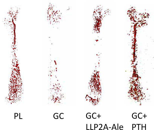
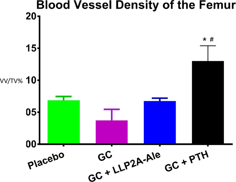
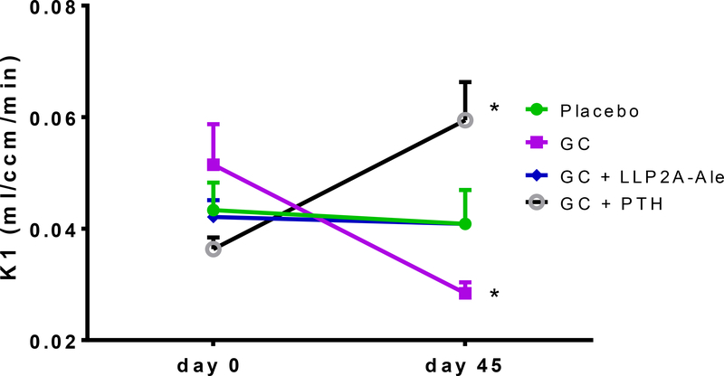
Recent findings: Glucocorticoids reduce osteoblast and osteocyte lifespan and activity and reduce the vascularity of the bone that together may explain the greater reductions in bone strength than those of bone mass. Treatments with parathyroid hormone fragments appear to reverse glucocorticoid-induced bone loss and fracture risk partially through maintaining bone vascularity and bone strength. This review identifies how glucocorticoid anti-osteogenic and vascular effects together may reduce bone strength. It also provides guidance to clinicians on rationale treatment for glucocorticoid-induced osteoporosis.

Keywords: Bone cells; Bone vascularity; Glucocorticoids; Osteonecrosis; Parathyroid hormone.


Conflict of interest statement

Compliance with Ethical Standards

Conflict of Interest

Nancy Lane reports having a patent (LLP2A-ale) issued.

Figures

Figure 1.
Figure 1.
Mice were treated with oral dexamethasone alone or with LLP2A-Ale (500ug/kg at day 0, 15, and 30) or PTH (1–34) (40ug/kg) 5 days/week for 45 days. Figure 1a. Images of blood vessels within the mouse femur At day 45, the mice had an intravenous infusion of Microfil (MV-120, Flow Tech, Inc., Carver, Massachusetts), and after perfusion, the animals were stored at 4°C overnight to allow polymerization. The following day, the femurs were dissected, fixed in 10% formalin, decalcified thoroughly and scanned using a micro-CT (VivaCT 40; Scanco Medical, Bassersdorf, Switzerland), with an isotropic resolution of 10.5 μm. The entire femur was used in the analysis for blood vessel density (VV)(14). Treatment with glucocorticoids appeared to reduce the bone vascularity while treatment with either LLP2A-Ale (500ug/kg at day 1, 14,28) or PTH (1–34) 40ug/kg 5x/wk maintained bone vascularity.
Figure 1b.
Figure 1b.
Blood vessel density of mouse femurs was reduced with glucocorticoid treatment and was maintained with either hPTH (1–34) or with a hybrid compound LLP2A-Ale An algorithm of thresholding 150–200, sigma 0.8 and support 1 were used for the quantification of the vascularization. Vascular density was presented as vessel volume per tissue volume (VV/TV) Treatment with glucocorticoid alone significantly reduced vascular density in the femur and treatment with either LLP2A-Ale (500ug/kg at day 1, 14,28 days) or PTH(1–34) (40ug/kg/5x wk) preserved the vascular density.
Figure 1c.
Figure 1c.
Bone Blood flow in the Proximal Tibia measured by 18F-fluoride by PET/CT. Mice were treated with oral dexamethasone alone or with LLP2A-Ale (500ug/kg at day1,14,28) or PTH (1–34) 5 days/week for 45 days. At day −1, and day 45 mice from each treatment group (n=6) had PET/CT scanning with the radiotracer 18F-fluoride to measure blood flow in vivo. Dynamic PET data were analyzed via a two-compartment, four-parameter model, and values of the rate constant K1 were compared between groups (Mean ± SD). At baseline, there were no differences in the K1 between the treatment groups. At day 45, compared to the placebo group, the GC group had a significant reduction in blood flow, and GC + PTH group had a significant increase compared to the placebo group.

References

    1. Weinstein RS. Glucocorticoid-induced osteoporosis and osteonecrosis. Endocrinology and metabolism clinics of North America. 2012;41:595–611. 10.1016/j.ecl.2012.04.004. - DOI - PMC - PubMed
    1. Steinbuch M, Youket TE, Cohen S. Oral glucocorticoid use is associated with an increased risk of fracture. Osteoporosis international: a journal established as result of cooperation between the European Foundation for Osteoporosis and the National Osteoporosis Foundation of the USA. 2004;15:323–328. 10.1007/s00198-003-1548-3. - DOI - PubMed
    1. Van Staa TP, Laan RF, Barton IP, Cohen S, Reid DM, Cooper C. Bone density threshold and other predictors of vertebral fracture in patients receiving oral glucocorticoid therapy. Arthritis Rheum. 2003. November;48(11):3224–9. 10.1002/art.11283. - DOI - PubMed
    1. Yao W, Cheng Z, Busse C, Pham A, Nakamura MC, Lane NE. Glucocorticoid excess in mice results in early activation of osteoclastogenesis and adipogenesis and prolonged suppression of osteogenesis: a longitudinal study of gene expression in bone tissue from glucocorticoid-treated mice. Arthritis Rheum. 2008;58:1674–1686. 10.1002/art.23954. - DOI - PMC - PubMed
    1. Yao W, Dai W, Jiang JX, Lane NE. Glucocorticoids and osteocyte autophagy. Bone. 2013. June;54(2):279–84. 10.1016/j.bone.2013.01.034. - DOI - PMC - PubMed

MeSH terms