Objective Assessment of Arterial Steal Phenomenon in Direct Carotid Cavernous Fistula Using 2D Parametric Parenchymal Blood Flow Analysis
- PMID: 30685958
- PMCID: PMC6433195
- DOI: 10.5469/neuroint.2018.01102
Objective Assessment of Arterial Steal Phenomenon in Direct Carotid Cavernous Fistula Using 2D Parametric Parenchymal Blood Flow Analysis
Abstract
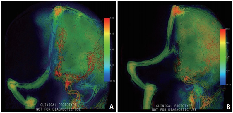
The aim of the study is to evaluate the hemodynamic changes and the parenchymal perfusion associated with carotid cavernous fistulas before and after embolization using two-dimensional (2D) parenchymal blood flow analysis. A 15-year-old boy presented with 2-month history of progressive right eye proptosis, chemosis, and diplopia after a motor vehicle accident. Intracranial liquid embolization using Onyx-18 through the inferior petrosal approach was done with balloon protection at the opening of the fistula in the internal carotid artery, resulting in complete occlusion of the fistula. Parenchymal blood flow analysis was done before and immediately after embolization. 2D parametric parenchymal blood flow analysis is newly introduced software that can provide data cannot be conveyed by conventional digital subtraction angiography alone. The software allows for objective assessment of the arterial steal and the parenchymal perfusion both pre, and post-embolization. Pre-embolization assessment may influence the therapeutic decision, while post-embolization assessment can evaluate the treatment efficacy.
Keywords: Embolization, Therapeutic; Fistula; Hemodynamics; Internal carotid artery; Software; Steal phenomenon.
Figures


Similar articles
-
Transvenous embolization with a combination of detachable coils and Onyx for a complicated cavernous dural arteriovenous fistula.Chin Med J (Engl). 2008 Sep 5;121(17):1651-5. Chin Med J (Engl). 2008. PMID: 19024093
-
Endovascular treatment of a direct post-traumatic carotid-cavernous fistula with electrolytically detachable coils.Wien Klin Wochenschr. 2006;118 Suppl 2:80-4. doi: 10.1007/s00508-006-0541-1. Wien Klin Wochenschr. 2006. PMID: 16817051
-
Surgical removal of embolic material after its unexpected migration through extracranial-intracranial anastomosis in the treatment of Barrow Type D carotid-cavernous fistula: case report.J Neurosurg. 2018 Mar;128(3):731-734. doi: 10.3171/2016.9.JNS152677. Epub 2017 Mar 3. J Neurosurg. 2018. PMID: 28298038
-
Endovascular techniques for treatment of carotid-cavernous fistula.J Neuroophthalmol. 2009 Mar;29(1):62-71. doi: 10.1097/WNO.0b013e3181989fc0. J Neuroophthalmol. 2009. PMID: 19458580 Review.
-
Bilateral carotid cavernous sinus fistula: a case report and review of the literature.J Neurol. 2018 Mar;265(3):453-459. doi: 10.1007/s00415-017-8657-y. Epub 2017 Nov 2. J Neurol. 2018. PMID: 29098418 Review.
References
-
- Costa VP, Molnar LJ, Cerri GG. Diagnosing and monitoring carotid cavernous fistulas with color Doppler imaging. J Clin Ultrasound. 1997;25:448–452. - PubMed
-
- Barrow DL, Spector RH, Braun IF, Landman JA, Tindall SC, Tindall GT. Classification and treatment of spontaneous carotid-cavernous sinus fistulas. J Neurosurg. 1985;62:248–256. - PubMed
-
- Chen YW, Jeng JS, Liu HM, Hwang BS, Lin WH, Yip PK. Carotid and transcranial color-coded duplex sonography in different types of carotid-cavernous fistula. Stroke. 2000;31:701–706. - PubMed
-
- Yanik B, Conkbayir I, Oztürk M, Acaroglu G, Hekimoglu B. Partial steal phenomenon in the ophthalmic artery due to a direct carotid-cavernous sinus fistula: orbital color Doppler ultrasonographic findings. J Ultrasound Med. 2003;22:1107–1110. - PubMed
LinkOut - more resources
Full Text Sources