. 2019 Feb 21;176(5):1128-1142.e18.
doi: 10.1016/j.cell.2018.12.023. Epub 2019 Jan 24.

A Unique Collateral Artery Development Program Promotes Neonatal Heart Regeneration

Affiliations

A Unique Collateral Artery Development Program Promotes Neonatal Heart Regeneration

Soumyashree Das et al. Cell. .

Abstract

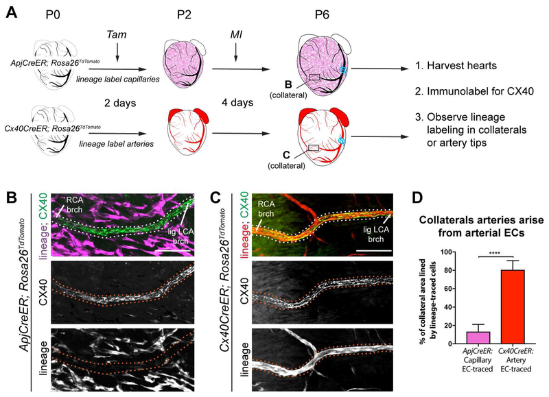
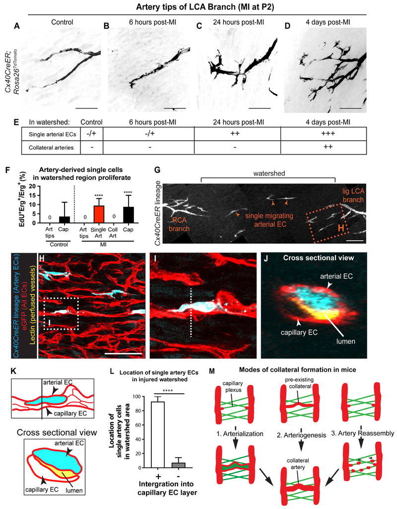
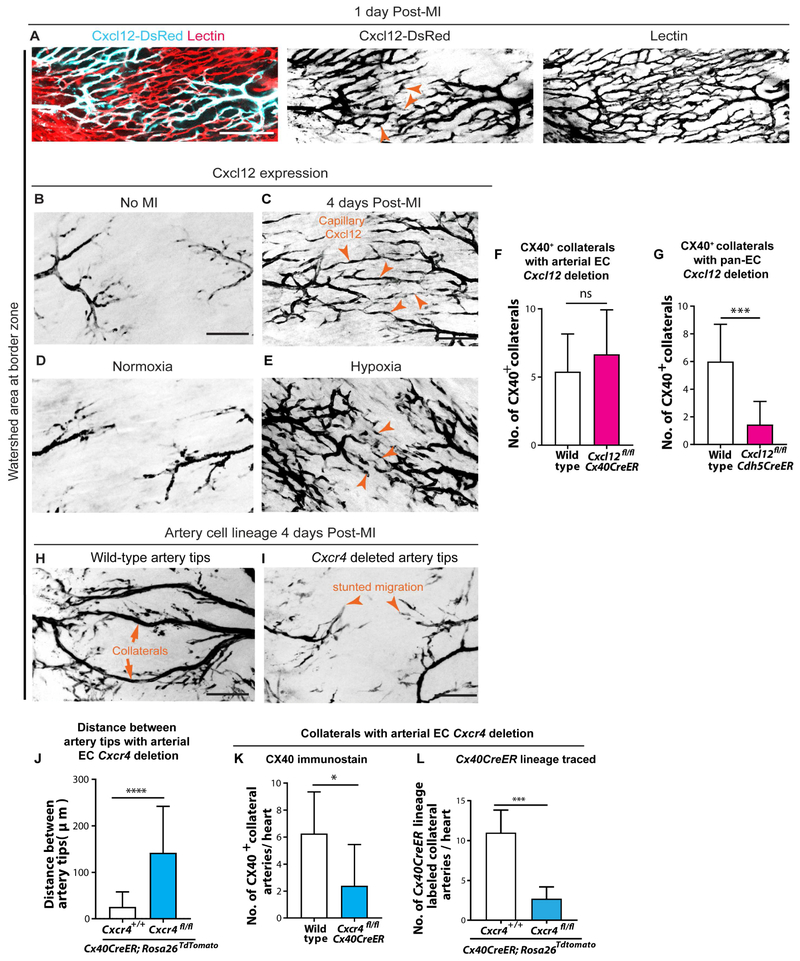
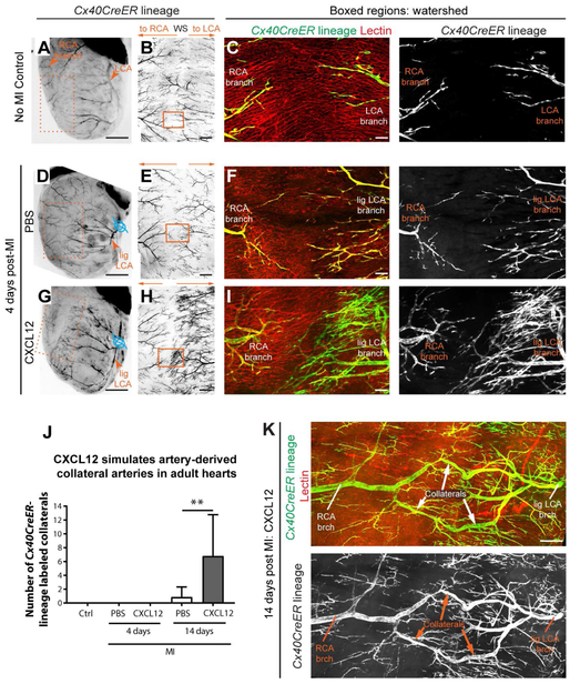
Collateral arteries are an uncommon vessel subtype that can provide alternate blood flow to preserve tissue following vascular occlusion. Some patients with heart disease develop collateral coronary arteries, and this correlates with increased survival. However, it is not known how these collaterals develop or how to stimulate them. We demonstrate that neonatal mouse hearts use a novel mechanism to build collateral arteries in response to injury. Arterial endothelial cells (ECs) migrated away from arteries along existing capillaries and reassembled into collateral arteries, which we termed "artery reassembly". Artery ECs expressed CXCR4, and following injury, capillary ECs induced its ligand, CXCL12. CXCL12 or CXCR4 deletion impaired collateral artery formation and neonatal heart regeneration. Artery reassembly was nearly absent in adults but was induced by exogenous CXCL12. Thus, understanding neonatal regenerative mechanisms can identify pathways that restore these processes in adults and identify potentially translatable therapeutic strategies for ischemic heart disease.

Keywords: CXCL12; arterialization; arteriogenesis; collateral arteries; endothelial cells; heart regeneration; myocardial infarction.


Figures

Figure 1.
Figure 1.. Extensive collateral artery formation in the neonatal mouse heart 4 days post MI. See also Figure S1 and S2.
(A) Experimental design where Cx40CreER-labeled arteries are red.(B) Confocal image of a watershed area from P2 control heart. Capillary bed (cyan) is fed by left (LCA) and right (RCA) coronary artery branches. (C and D) Confocal images (anterior views) of control (C) and myocardial infarction (MI) (D) hearts. Arterial ECs are black. MI induced collateral arteries that connect ligated (lig) branches (brchs) of LCA with RCA branches across the watershed area (boxed regions). Asterisks are RCA showing through from the posterior heart wall. (E) Schematic of how collateral arteries restore blood flow (arrows) to injured myocardium. Arteries, red; ligation, blue. (F) Some Cx40CreER-labeled collateral arteries contain smooth muscle. (G and H) Quantification of Cx40CreER-labeled (G) and smooth muscle covered (H) collateral arteries. Hearts: n=8 control, n=12 MI. (I) Collateral arteries were similar in diameter to quaternary (4֯) branches . Arteries: N=24 CX40+, n=9 αSMA+. (J) Collateral arteries were perfused. Cap, capillaries; P, postnatal; Tam, Tamoxifen; EC, endothelial cells; TdTom, TdTomato. Scale bars: B, 200μm; C, D low mag, 500μm; C, D boxed region, 200μm; F and J, 200μm. Error bars are st dev: ***, p≤0.001; ****, p≤0.0001.
Figure 2.
Figure 2.. Collateral arteries are formed by arterial endothelial cells. See also Figure S3.
(A) Experimental design where non-labeled arteries are black and lineage-labeled capillaries or arteries are magenta or red, respectively. Boxes localize collateral arteries in B and C. (B and C) CX40+ collateral arteries (dotted lines) showed minimal ApjCreER lineage labeling from capillaries (B), but heavy lineage-labeling Cx40CreER+ arteries (C). (D) Quantification of B and C (n=7 arteries each). Lig LCA, ligated left coronary artery; MI, myocardial infarction; RCA, right coronary artery; Tam, Tamoxifen; P, postnatal; EC, endothelial cells; brch, branch. Scale bars: 100μm. Error bars are st dev: ****, p≤0.0001.
Figure 3:
Figure 3:. Artery-derived collateral development is restricted to the neonatal regenerative window.
(A) Anterior view of P6 Cx40CreER; Rosa26TdTomato heart. Tamoxifen at P0, MI at P2, and harvested 4 days post-MI. Arteries are black, ligation in blue. Asterisks, right coronary artery (RCA) from posterior heart wall. (B) Boxed region from A. Arrowheads, collateral artery spanning watershed region. (C) Boxed region from B with non-coalesced arterial ECs in space between ligated left coronary artery (lig LCA) and non-ligated RCA branches. Pattern seen in n=8 hearts. (D) Anterior view of P11 Cx40CreER; Rosa26TdTomato. Tamoxifen at P0, ligated at P7, and harvested 4 days post-MI. (E) Boxed region from D reveal few lineage labeled artery cells in watershed and no collateral arteries. (F) Quantification at 4 days post-MI. Hearts: P2, n=12; P7, n=6. (G) Lectin perfusion in healthy and infarcted heart regions injured at P2 or P7, 4 days post-MI. (H) Quantification of mean fluorescence intensity. Hearts: P7, n=6, P2 n=6. Brch, branch; P, postnatal; EC, endothelial cells; MI, myocardial infarction. Scale bars: A and D, 500μm; B and E, 200 μm; G, 100μm. Error bars are st dev: *, p≤0.05; ****, p≤0.0001.
Figure 4.
Figure 4.. Migration of individual arterial ECs precedes the appearance of artery-derived collaterals. See also Figure S4.
(A-D) LCA tips from control or injured hearts. Arterial ECs appear mobilized within 24 hours. Tamoxifen injected at P0; MI at P2. Pattern seen in n=6 control and n=4 MI hearts each. (E) Table summarizing single arterial ECs in watersheds before collateral artery development. –, absent; −/+, very few; ++, many; +++, very many. (F) EdU incorporation indicates that artery cells in watershed proliferate. Hearts: n=2 control, n=3 MI. (G-J) Cx40CreER; Rosa26TdTomato; Tie2;:eGFP::Claudin5 hearts subjected to MI at P2 and Lectin perfused before isolation at P6. (G) Single artery cells (arrowheads) in watershed area 4 days post-MI. (H) Box in G showing arterial ECs (cyan) closely associated with capillaries (red). (I) Box in H. (J) Cross-section view (dotted line in I). Artery cell is adjacent to perfused Lectin (yellow), indicating it’s within EC layer. (K) Schematic of I and J. EC (L) Quantification where single artery cells are within EC layer (n=5 hearts). (M) Schematic adding Artery Reassembly to the different modes of collateral artery formation. LCA, left coronary artery; MI, myocardial infarction; P, postnatal; Cap, capillary; Coll, collateral artery; Art, artery. Scale bars:100μm. Error bars are st dev: ****, p≤0.0001.
Figure 5.
Figure 5.. CXCR4 signaling guides arterial migration during collateral formation. See also Figure S5 and S6.
(A) Cxcl12-DsRed expression in watershed capillaries 1 day post-myocardial infarction (MI). Pattern seen in 4 hearts. (B-E) Cxcl12-DsRed in P6 hearts. Cxcl12 is in artery tips in non-injured hearts (B), but is induced in capillary ECs in watershed upon MI (arrowheads, C). Pattern seen in n=4 control and n=6 MI hearts. In comparison to normoxic conditions (D), hypoxia expands Cxcl12-DsRed into capillary ECs (arrowheads, E). Pattern seen in n=4 normoxic hearts and n=13 hypoxic hearts. (F and G) CX40+ collaterals in injured P6 hearts following arterial Cxcl12 deletion (F) (n=10 wild-type, n=6 knockout (KO) hearts) or pan-EC Cxcl12 deletion (G) ECs (n=14 wild-type, n=9 KO hearts). (H and I) Cx40CreER-lineage labeled artery cells and collaterals (H) in watershed are dramatically decreased with EC arterial Cxcr4 deletion (I). (J-L) Number of single arterial ECs in watershed (J) (n=3 wild-type, n=4 KO hearts), CX40+ collaterals (K) (n=2 wild-type, n=6 KO hearts), and Cx40CreER lineage-traced collaterals (L) (n=11 wild-type, n=5 KO hearts) is significantly reduced upon arterial deletion of Cxcr4. All deletions induced at P0; MIs at P2. lig, ligated; LCA, left coronary artery; RCA, right coronary artery; P, postnatal; EC, endothelial cell. Scale bars: 100μm. Error bars are st dev: *, p≤0.05; ***, p≤0.001, ****, p≤0.0001.
Figure 6.
Figure 6.. Endothelial Cxcl12 and Cxcr4 facilitate neonatal heart regeneration. See also Figure S5 and S6.
(A) Model for collateral formation in neonates upon MI. (B-E) Ejection fractions 28 days post-MI. No decrease in controls (B) (n=8 No-MI, n=14 MI animals). Function is reduced with Pan-EC Cxcl12 deletion (C) (n=12 No-MI, n=9 MI animals) and arterial Cxcr4 deletion (D) (n=8 No-MI, n=8 MI animals) 28 days post-MI. (E) Normal function in non-injured hearts with postnatal endothelial gene deletions. N= 8 controls, n=12 Cxcl12fl/fl; Cdh5CreER, n=8 Cxcr4fl/fl; Cx40CreER animals. (F) Compared to controls (n=12), fibrotic scar was increased in endothelial Cxcl12 (n=11 hearts) and arterial Cxcr4 (n=7 hearts) knockouts. (G) Transverse section indicating ischemic zone, border zone and remote zone. (H) Reduced PH3+ cardiomyocytes in ischemic, border, and remote zone of neonatal hearts lacking endothelial Cxcl12 (n≥5) or arterial Cxcr4 (n=5), 7 days post-MI. lig, ligated; LCA, left coronary artery; RCA, right coronary artery; P, postnatal; Endo, endothelial; EC, endothelial cell; MI, myocardial infarction. Error bars are st dev: *, p≤0.05; **, p≤0.01; ***, p≤0.001, ****, p≤0.0001.
Figure 7.
Figure 7.. CXCL12 administration induces collateral formation through Artery Reassembly in adult hearts. See also Figure S7.
(A, D, and G) Stereoscope images of adult Cx40CreER; Rosa26TdTomato hearts. Arterial endothelial cells, black; ligation, blue. Tamoxifen given 2 days before MI; heart isolated 4 days post-MI. (B, E, and H) Confocal imaging of isolated anterior heart walls (boxes in A, D, and G) showing left (LCA) and right (RCA) coronary artery branches in watershed (WS). (C, F, and I) Boxed regions in B, E, and H showing Cx40CreER-lineage and perfused Lectin. CXCL12 increases artery cell outgrowth from LCA branches. (J) CXCL12 treatment induced artery-derived collaterals at 14 days post-MI (from left to right: n=4, n=5, n=10, n=13, and n=14 hearts). (K) Artery-derived collateral from CXCL12 administered heart Ctrl, control; Lig, ligated; MI, myocardial infarction; brch, branch. Scale bars: A, D, and G, 2 mm; B, E, and H, 500μm; C, F, and I, 100μm; K, 200μm. Error bars are st dev: **, p≤0.01.

Comment in

References

    1. Abbott JD, Huang Y, Liu D, Hickey R, Krause DS, and Giordano FJ (2004). Stromal cell-derived factor-1alpha plays a critical role in stem cell recruitment to the heart after myocardial infarction but is not sufficient to induce homing in the absence of injury. Circulation 110, 3300–3305. - PubMed
    1. Askari AT, Unzek S, Popovic ZB, Goldman CK, Forudi F, Kiedrowski M, Rovner A, Ellis SG, Thomas JD, DiCorleto PE, et al. (2003). Effect of stromal-cell-derived factor 1 on stem-cell homing and tissue regeneration in ischaemic cardiomyopathy. Lancet 362, 697–703. - PubMed
    1. Aurora AB, Porrello ER, Tan W, Mahmoud AI, Hill JA, Bassel-Duby R, Sadek HA, and Olson EN (2014). Macrophages are required for neonatal heart regeneration. J. Clin. Invest 124, 1382–1392. - PMC - PubMed
    1. Bassat E, Mutlak YE, Genzelinakh A, Shadrin IY, Baruch Umansky K, Yifa O, Kain D, Rajchman D, Leach J, Riabov Bassat D, et al. (2017). The extracellular matrix protein agrin promotes heart regeneration in mice. Nature 547, 179–184. - PMC - PubMed
    1. Baxter R, Hastings N, Law A, and Glass EJ. (2008). CXCL12 gene expression is upregulated by hypoxia and growth arrest but not by inflammatory cytokines in rheumatoid synovial fibroblasts. Anim. Genet 39, 561–563.

Publication types

LinkOut - more resources