. 2019 Feb 6;101(3):412-420.e3.
doi: 10.1016/j.neuron.2019.01.012. Epub 2019 Jan 24.

A Mast-Cell-Specific Receptor Mediates Neurogenic Inflammation and Pain

Affiliations

A Mast-Cell-Specific Receptor Mediates Neurogenic Inflammation and Pain

Dustin P Green et al. Neuron. .

Abstract

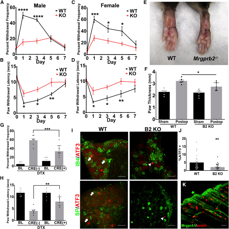
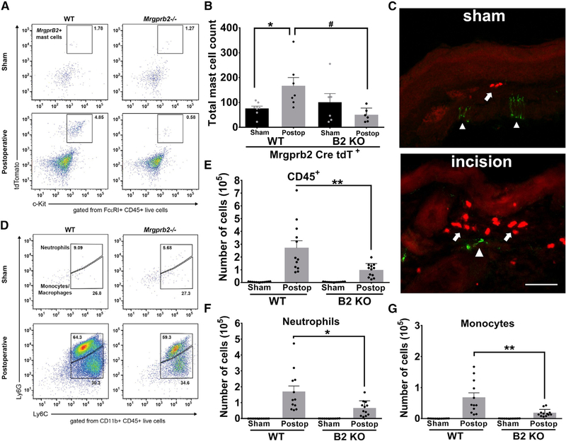
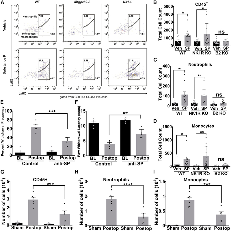
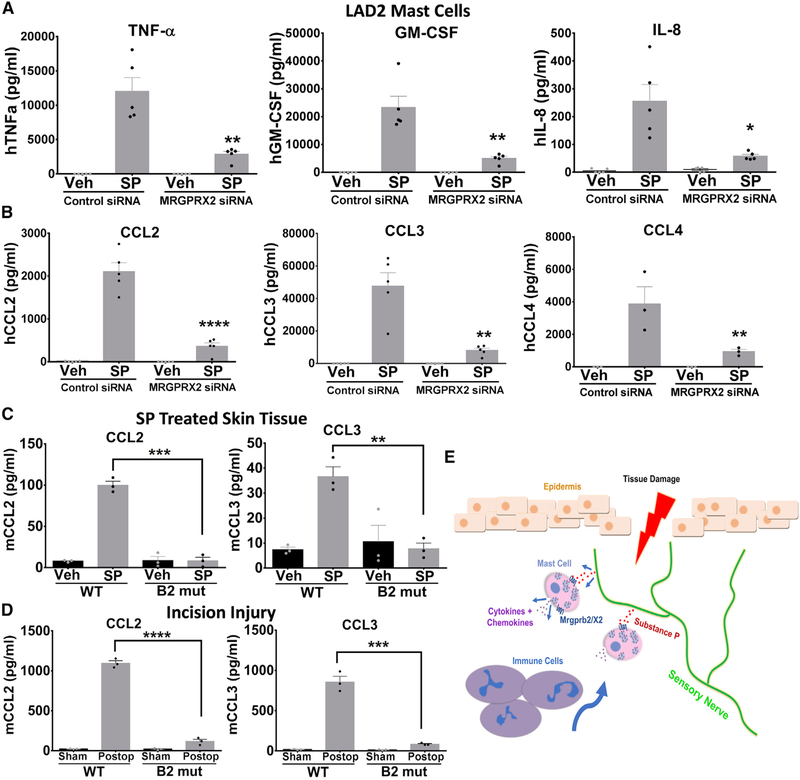
Mast cells can be found in close proximity to peripheral nerve endings where, upon activation, they release a broad range of pro-inflammatory cytokines and chemokines. However, the precise mechanism underlying this so-called neurogenic inflammation and associated pain has remained elusive. Here we report that the mast-cell-specific receptor Mrgprb2 mediates inflammatory mechanical and thermal hyperalgesia and is required for recruitment of innate immune cells at the injury site. We also found that the neuropeptide substance P (SP), an endogenous agonist of Mrgprb2, facilitates immune cells' migration via Mrgprb2. Furthermore, SP activation of the human mast cell led to the release of multiple pro-inflammatory cytokines and chemokines via the human homolog MRGPRX2. Surprisingly, the SP-mediated inflammatory responses were independent of its canonical receptor, neurokinin-1 receptor (NK-1R). These results identify Mrgprb2/X2 as an important neuroimmune modulator and a potential target for treating inflammatory pain.


Conflict of interest statement

DECLARATION OF INTERESTS

The authors declare no competing interest.

Figures

Figure 1.
Figure 1.. Mrgprb2−/− Mice Exhibit Significantly Less Inflammation-Induced Hypersensitivity in an Incision Model of Inflammatory Pain
(A–D) An incision-induced model of postoperative pain was performed on WT or Mrgprb2−/− mice. Mechanical (A) and thermal (B) hypersensitivity were measured starting 24 h after incision for up to 7 days. (BL, baseline, no difference was observed in baseline between WT and Mrgprb2−/− mice.) Female mice were also tested for mechanical (C) and thermal (D) hypersensitivity starting 24 h after incision for up to 7 days. (E) Representative images of postoperative WT and Mrgprb2−/− hindpaws 24 h after incision. (F) Quantification of incision-induced inflammation, measured by paw thickness, in WT and Mrgprb2−/− mice. (G and H) Mechanical (G) and thermal (H) hypersensitivity was measured 24 h after incision in DTX-treated Mrgprb2Cre(−) DTR and Mrgprb2Cre(+) DTR mice. (I) Confocal microscopy immunofluorescence images of DRG sections comparing WT to Mrgprb2−/− (B2 KO) mice in the incision model of postoperative pain. In the top panel, arrows point to ATF-positive nuclei (red) co-stained with IB4 (green). The bottom panel arrows point to ATF-positive nuclei (red) co-stained with substance P (SP, green). Scale bars indicate 50 μm. (J) Graph of percentage of ATF+ DRG nuclei comparing postoperative WT and Mrgprb2−/− mice. (K) Confocal image of a hindpaw skin section stained with anti-GFP antibody (green) to visualize MrgprA1-GFP expressing nerve fibers in the dermis. The section was counterstained with Avidin to label mast cells. Scale bar indicates 50 μm. Data were analyzed using two-tailed Student’s t test and two-way ANOVA with Bonferroni’s post hoc test (> 2 groups); *p < 0.05, **p<0.01, ****p < 0.001, n = 6–7/group; error bar, SEM.
Figure 2.
Figure 2.. Involvement of Mrgprb2 in Immune Cell Recruitment in a Postoperative Model of Inflammatory Pain
(A) Representative flow cytometric profiles of biopsies taken from Mrgprb2-Cre tdT WT or Mrgprb2-Cre tdT Mrgprb2−/− hindpaw skin collected from sham or incision-injured animals. Numbers indicate the percentage of cells pre-gated on viability, CD45+, FcεRI+. (B) The absolute number of Mrgprb2-Cre tdT + mast cells was calculated from the flow cytometric profile for each mouse. (C) Confocal image of mouse hindpaw sections comparing sham to incision. Mrgprb2-Cre tdT + mast cells (red) are identified with arrows. Peripheral afferents (green) are stained with Neurofilament 200 and are identified with arrowheads. Scale bars indicate 100 μm. (D) Representative flow cytometric profiles of biopsies taken from WT or Mrgprb2−/− hindpaw skin 24 h after incision injury. Numbers indicate the percentage of cells pre-gated on viability, CD45+, CD11b+. (E-G) The absolute number of cells are shown for CD45+ cells (E), CD11b+Ly6G+ neutrophils (F), or CD11b+Ly6G-Ly6C+ monocytes (G). Data were analyzed using two-tailed Student’s t test. *p < 0.05, **p < 0.01, n = 11/group; error bar, SEM.
Figure 3.
Figure 3.. Substance P Promotes Innate Immune Cell Recruitment via Mrgprb2
(A) Representative flow cytometric profiles of biopsies taken from WT, NK-1R−/− or Mrgprb2−/− hindpaw skin injected with vehicle or substance P (50 μM in 10 μL). Numbers indicate the percentage of cells pre-gated on viability, CD45+, CD11b+. (B-D) The absolute number of each immune cell was calculated from the flow cytometric profile for each mouse and is shown for CD45+ cells (B), CD11b+Ly6G+ neutrophils (C), or CD11b+Ly6G-Ly6C+ monocytes (D). (E and F) Anti-substance-P antibodies (15 μg) or vehicle were injected into the hindpaws of WT and Mrgprb2−/− mice prior to incision-induced injury. At 23 h after incision, anti-substance-P antibodies (15 μg) were injected again, and mechanical (E) and thermal (F) hypersensitivity were measured 1 h later (BL, baseline). (G-I) Biopsies taken from WT hindpaw skin treated with anti-substance-P or isotype control antibodies 24 h after incision injury. The absolute number of cells are shown for CD45+ cells (G), CD11b+Ly6G+ neutrophils (H), or CD11b+Ly6G-Ly6C+ monocytes (I). Data were analyzed using two-tailed Student’s t test. *p < 0.05, **p < 0.01, n = 13/group for WT, NK-1R−/−, and Mrgprb2−/− flow cytometry, n = 5/group for behavior, n = 6/group for flow analysis of anti-SP-treated tissue; error bar, SEM.
Figure 4.
Figure 4.. Substance-P-Treated Human Mast Cells Release Inflammatory Cytokines and Chemokines via MRGPRX2 Activation
(A and B) LAD2 mast cells were treated with siRNA against MRGPRX2 or control siRNA. Concentrations of (A) cytokines TNF-a, GM-CSF, IL-8, and (B) chemokines CCL2, CCL3, and CCL4 in supernatant taken from vehicle-treated or substance-P-treated (1 μM) LAD2 mast cells were determined by ELISA. (C) Hindpaw tissue injected with vehicle or substance P (50 μM) was biopsied 24 h later, and CCL2 and CCL3 levels were determined by ELISA. (D) 24 h after incision injury, hindpaw tissue was biopsied, and CCL2 and CCL3 levels were determined by ELISA. (E) Diagram depicting the mechanism by which tissue damage and neuronal release of substance P activates MrgprX2/b2, leading to the release of cytokines and the subsequent recruitment of immune cells to the site of injury. Data were analyzed using a one-way ANOVA. *p < 0.05, **p < 0.01, ****p < 0.001; error bar, SEM.

Comment in

References

    1. Abbadie C, Lindia JA, Cumiskey AM, Peterson LB, Mudgett JS, Bayne EK, DeMartino JA, MacIntyre DE, and Forrest MJ (2003). Impaired neuropathic pain responses in mice lacking the chemokine receptor CCR2. Proc. Natl. Acad. Sci. USA 700, 7947–7952. - PMC - PubMed
    1. Azimi E, Reddy VB, Shade KC, Anthony RM, Talbot S, Pereira PJ, and Lerner EA (2016). Dual action of neurokinin-1 antagonists on Mas-related GPCRs. JCI Insight 7, e89362. - PMC - PubMed
    1. Azimi E, Reddy VB, Pereira PJS, Talbot S, Woolf CJ, and Lerner EA (2017). Substance P activates Mas-related G protein-coupled receptors to induce itch. J. Allergy Clin. Immunol 740, 447–453.e3. - PMC - PubMed
    1. Barabas ME, and Stucky CL (2013). TRPV1, but notTRPA1, in primary sensory neurons contributes to cutaneous incision-mediated hypersensitivity. Mol. Pain 9, 9. - PMC - PubMed
    1. Chatterjea D, Wetzel A, Mack M, Engblom C, Allen J, Mora-Solano C, Paredes L, Balsells E, and Martinov T (2012). Mast cell degranulation mediates compound 48/80-induced hyperalgesia in mice. Biochem. Biophys. Res. Commun 425, 237–243. - PMC - PubMed

Publication types