. 2019 Feb 4;29(3):449-460.e2.
doi: 10.1016/j.cub.2018.12.043. Epub 2019 Jan 24.

The DNA Damage Checkpoint and the Spindle Position Checkpoint Maintain Meiotic Commitment in Saccharomyces cerevisiae

Affiliations

The DNA Damage Checkpoint and the Spindle Position Checkpoint Maintain Meiotic Commitment in Saccharomyces cerevisiae

Olivia Ballew et al. Curr Biol. .

Abstract

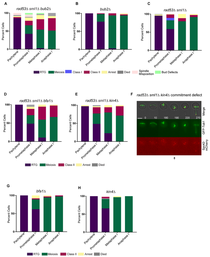
During meiosis, diploid progenitor cells undergo one round of DNA replication followed by two rounds of chromosome segregation to form haploid gametes. Once cells initiate the meiotic divisions, it is imperative that they finish meiosis. A failure to maintain meiosis can result in highly aberrant polyploid cells, which could lead to oncogenesis in the germline. How cells stay committed to finishing meiosis, even in the presence of a mitosis-inducing signal, is poorly understood. We addressed this question in budding yeast, in which cells enter meiosis when starved. If nutrient-rich medium is added before a defined commitment point in mid-prometaphase I, they can return to mitosis. Cells in stages beyond the commitment point will finish meiosis, even with nutrient addition. Because checkpoints are signaling pathways known to couple cell-cycle processes with one another, we asked if checkpoints could ensure meiotic commitment. We find that two checkpoints with well-defined functions in mitosis, the DNA damage checkpoint and the spindle position checkpoint, have crucial roles in meiotic commitment. With nutrient-rich medium addition at stages beyond the commitment point, cells that are deficient in both checkpoints because they lack Rad53 and either Bub2, Bfa1, or Kin4 can return to mitotic growth and go on to form polyploid cells. The results demonstrate that the two checkpoints prevent cells from exiting meiosis in the presence of a mitosis-inducing signal. This study reveals a previously unknown function for the DNA damage checkpoint and the spindle position checkpoint in maintaining meiotic commitment.


Conflict of interest statement

Declaration of Interests

The authors do not have any competing interests to declare.

Figures

Figure 1.
Figure 1.. The DNA damage checkpoint prevents polyploidy upon RTG from prometaphase I.
(A) Cartoon of meiotic commitment. (B, C) Representative time-lapse images of a cell committed to meiosis (B) and underwent RTG (C) after nutrient-rich medium addition in prometaphase I. (D, E) Graph of the percentage of WT cells (D) and mec1Δ sml1Δ cells (E) with indicated outcomes upon nutrient-rich medium addition at each meiotic stage (n = 308 (D) and n=293 (E)). Three independent experiments were performed for each strain. (F, G) Representative time-lapse images of a cell that displayed the class I (F) and class II (G) phenotype. (H, I) Plot of spindle length (μm) at time of nutrient-rich medium addition in prometaphase I of wildtype (H) and mec1Δ sml1Δ (I) cells. Cells were categorized by outcome and corresponding spindle length (n = 35 for each category). Error bars represent SEM (standard error of the mean). (J, K) Graph of the percentage of cells for each outcome upon nutrient-rich medium addition during prometaphase I in indicated mutants ((n ≥ 50 prometaphase I cells for each mutant (J) and n ≥ 35 prometaphase I cells for each mutant (K), three independent experiments for each strain)). Asterisks indicate a statistically significant difference (H, I p < 0.05, Mann-Whitney Test) (J, K p < 0.05 rx Contingency Table). In all time-lapses (B, C, F, G), Cells expressed GFP-Tub1 and Spc42-mCherry. Numbers indicate time (mins) from nutrient-rich medium addition at prometaphase I. Scale Bars: 5μm. Arrows indicate time of bud emergence. See also Figure S1.
Figure 2.
Figure 2.. Persistent DSBs cause a DNA damage checkpoint delay in meiosis II
(A, B) Representative time-lapse images of a wildtype (WT) cell with a Rad52-GFP focus that persisted throughout the meiotic divisions (A), or a Rad52-GFP focus that disappeared during prophase I (B). Cells also expressed Spc42-mCherry. Numbers indicate time (mins) relative to prometaphase I initiation (spindle pole body separation). Scale Bars: 5μm. (C) Graph of percentage of wildtype, mec1Δ sml1Δ, and spo11Δ cells in meiosis with Rad52-GFP foci in prometaphase I (n ≥ 48 cells for each). Three independent experiments performed for each strain. (D, E) Graph of the average time (mins) from prometaphase I to anaphase I (D), and anaphase I to anaphase II (E) in wildtype cells with or without Rad52-GFP foci in prometaphase I. Error bars represent SEM. (n ≥ 44 cells). Asterisks indicate a statistically significant difference (C, p < 0.0001, Fisher’s Exact Test) (D, E p < 0.05, Mann-Whitney test). ns represents not significant.
Figure 3.
Figure 3.. Cells with persistent Rad52-GFP that undergo RTG from prometaphase I have a DNA damage checkpoint delay
(A, B) Representative time-lapse images of a cell that underwent RTG (A), and finished meiosis (B) after nutrient-rich medium addition in prometaphase I. Cells expressed Rad52-GFP and Spc42-mCherry. Numbers indicate time (mins) from nutrient-rich medium addition in prometaphase I. Scale bars: 5μm. Arrow indicates time of bud emergence. (C) Graph of the percentage of WT and mec1Δ sml1Δ cells that underwent RTG from prometaphase I with Rad52-GFP foci (n ≥ 35 cells). Three independent experiments performed for each strain. (D-G) Graph of average time (mins) from nutrient-rich medium addition in prometaphase I to mitotic anaphase (D), bud formation (E), anaphase I (F), or from anaphase I to anaphase II (G) in WT and mec1Δ sml1Δ cells with Rad52-GFP foci in prometaphase I (n = 35). Error bars represent SEM. Asterisks indicate a statistically significant difference from WT (D-G, p < 0.05, Mann-Whitney Test) (C, p < 0.0001, Fisher’s Exact Test). ns represents not significant.
Figure 4.
Figure 4.. Class I and class II mec1Δ sml1Δ cells segregate homologous chromosomes in the first division
(A-D) Representative time-lapse images and cartoons of a cell that underwent RTG (A) finished meiosis (B) displayed the class I (C) and class II phenotype (D) ((n=50 cells for (A) and (B) and n=10 cells for (C) and (D)). Cells expressed LacI-GFP and Spc42-mCherry. Numbers indicate time (mins) from nutrient-rich medium addition at prometaphase I. Scale bars: 5μm. Arrows indicate time of bud emergence.
Figure 5.
Figure 5.. The spindle position checkpoint delays Cdc14 release in Class I cells.
(A-D) Representative time-lapse images of a cell that underwent RTG (A), finished meiosis (B), displayed the class I phenotype (C), and class II phenotype (D) upon nutrient-rich medium addition in prometaphase I. Cells expressed Cdc14-GFP and Spc42-mCherry. Numbers indicate time (mins) from nutrient-rich medium addition at prometaphase I. Scale bars: 5μm. Arrows indicate time of bud emergence. See also Figure S2.
Figure 6.
Figure 6.. Cells fail to stay committed to meiosis in the absence of both the DNA damage checkpoint and spindle position checkpoint.
(A-E, G, H) Graph of percentage of rad53Δ sml1Δ bub2Δ (A), bub2Δ (B), rad53Δ sml1Δ (C), rad53Δ sml1Δ bfa1Δ (D), and rad53Δ sml1Δ kin4Δ (E), bfa1Δ (G), and kin4Δ cells (H), with indicated outcomes upon nutrient-rich medium addition at each meiotic stage ((n= 152 (A), n=157 (B), n=216 (C), n=149 (D), n=140 (E), n=145 (G) and n=158 (H)). Two or more independent experiments performed for each strain. (rad53Δ sml1Δ kin4Δ, rad53Δ sml1Δ bfa1Δ, and rad53Δ sml1Δ bub2Δ all displayed a statistically significant difference from wildtype in prometaphase I, metaphase I, and anaphase I. The above mutants were also significantly different from the corresponding single mutant (kin4Δ, bfa1Δ, and bub2Δ, respectively) in prometaphase I, metaphase I, and anaphase I (p < 0.01, rx Contingency Tables)). (F) Representative time-lapse images of a rad53Δ sml1Δ kin4Δ cell expressing GFP-Tub1 and Spc42-mCherry that is uncommitted. Numbers indicate time (mins) from nutrient-rich medium addition at anaphase I. Scale bar: 5mm. Arrow indicates time of bud emergence. See also Figure S3.

References

    1. Winter E (2012). The Sum1/Ndt80 transcriptional switch and commitment to meiosis in Saccharomyces cerevisiae. Microbiol Mol Biol Rev 76, 1–15. - PMC - PubMed
    1. Page AW, and Orr-Weaver TL (1997). Stopping and starting the meiotic cell cycle. Curr Opin Genet Dev 7, 23–31. - PubMed
    1. Nebreda AR, and Ferby I (2000). Regulation of the meiotic cell cycle in oocytes. Curr Opin Cell Biol 12, 666–675. - PubMed
    1. Kimble J (2011). Molecular regulation of the mitosis/meiosis decision in multicellular organisms. Cold Spring Harb Perspect Biol 3, a002683. - PMC - PubMed
    1. Sherman F, and Roman H (1963). Evidence for two types of allelic recombination in yeast. Genetics 48, 255–261. - PMC - PubMed

Publication types

MeSH terms