. 2019 Jan 27;11(2):724-740.
doi: 10.18632/aging.101774.

CD56-negative NK cells with impaired effector function expand in CMV and EBV co-infected healthy donors with age

Affiliations

CD56-negative NK cells with impaired effector function expand in CMV and EBV co-infected healthy donors with age

Bojana Müller-Durovic et al. Aging (Albany NY). .

Abstract

Natural killer cells lacking expression of CD56 (CD56neg NK cells) have been described in chronic HIV and hepatitis C virus infection. Features and functions of CD56neg NK cells in the context of latent infection with CMV and / or EBV with age are not known. In a cohort of healthy donors >60 years of age, we found that co-infection with CMV and EBV drives expansion of CD56neg NK cells. Functionally, CD56neg NK cells displayed reduced cytotoxic capacity and IFN-γ production, a feature that was enhanced with CMV / EBV co-infection. Further, the frequency of CD56neg NK cells correlated with accumulation of end-stage-differentiated T cells and a reduced CD4 / CD8 T cell ratio, reflecting an immune risk profile. CD56neg NK cells had a mature phenotype characterized by low CD57 and KIR expression and lacked characteristics of cell senescence. No changes in their activating NK cell receptor expression, and no upregulation of the negative co-stimulation receptors PD-1 or TIM-3 were observed. In all, our data identify expansion of dysfunctional CD56neg NK cells in CMV+EBV+ elderly individuals suggesting that these cells may function as shape-shifters of cellular immunity and argue for a previously unrecognized role of EBV in mediating immune risk in the elderly.

Keywords: CD56-negative; CMV; EBV; NK cells; aging; exhaustion; senescence.


Conflict of interest statement

CONFLICTS OF INTEREST: All authors declare no competing financial interests.

Figures

Figure 1
Figure 1
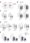
CD56neg NK cells with impaired effector function expand in CMV and EBV co-infected individuals >60 years of age. (A) Frequencies of CD56bright, CD56dim and CD56neg NK cells in YOUNG (<35 years) CMV (gray bars, n=10/10) and CMV+ (black bars, n=10/10) individuals compared to OLD (>60 years) CMV- (gray bars, n=20/21) and CMV+ (black bars, n=17/20) donors analyzed as in Supplementary Figure S1A. (B) Representative FACS dot plots from a CMVEBV and a CMV+EBV+ donor are shown. Numbers indicate the percentage of cells within total NK cells in peripheral blood. (C) Absolute cell numbers for CD56dim and CD56neg NK cells – as determined by FACS analysis in total PBMCs– are shown in a cohort of HDs >60 years of age stratified as CMVEBV (n=11/11), CMVEBV+ (n=10/24), CMV+EBV (n=6/6), and CMV+EBV+ (n=12/14). (D-F) FACS-sorted CD56dim and CD56neg NK cells from CMVEBV (n=7), CMVEBV+ (n=4), CMV+EBV- (n=4) and CMV+EBV+ (n=5) donors were either left un-stimulated (empty bars), stimulated with IL-12 / IL-18 (green bars) or K562 target cells (blue bars) and (D) CD107a expression (E) target cell lysis and (F) IFN-γ production were assessed after 6 hours of (co-)culture. Parametric data were compared by Student’s t-test and are shown as mean ± SEM, non-parametric data by Mann-Whitney test and are shown as median ± IQR, respectively. * p≤0.05, ** p≤0.005, *** p≤0.005.
Figure 2
Figure 2
CD56neg NK cells do not acquire cell senescence characteristics. (A) Frequencies of CD56neg NK cells in relation to CD8+ EMRA T cells (left panel), CD27-CD28- T cells (middle panel) and the CD4 / CD8 T cell ratio (right panel) as assessed by FACS analysis in total PBMCs (n=53/55). Data were analyzed by linear regression: correlation strength (R2) and statistical significance (p-value) are indicated for each scatter plot. (B) The differentiation stage of CD56neg NK cells was assessed by FACS analysis for NKG2A, CD62L, KIR and CD57 expression in total PBMCs. CD56neg NK cells were compared to CD56bright and CD56dim NK cells from CMVEBV (gray bars, n=10/11) and CMV+EBV+ donors (black bars, n=10/14). (C) Proliferation of NK cell subsets from CMVEBV (gray bars, n=10/11) and CMV+EBV+ (black bars, n=10/14) donors as assessed directly ex vivo by FACS analysis for Ki-67 expression. (D) Telomere length of NK cell subsets in CMVEBV (gray bars, n=10/10) and CMV+EBV+ donors (black bars, n=10/14) as assessed by FACS-based FISH-technique. Data are shown as geometric mean of fluorescence intensity (gMFI) of the telomere probe (TelC), normalized to the gMFI TelC value of the total lymphocyte population for each donor. (E) Global phosphorylation of the histone H2A.X (γH2A.X Ser139) in CD56dim and CD56neg NK cells in CMVEBV (gray bars, n=8/11) and CMV+EBV+ donors (black bars, n=9/14) as assessed directly ex vivo by FACS analysis. (F) Representative histograms for γH2A.X staining in a CMVEBV (gray histograms) and CMV+EBV+ (blue histograms) donor. UV-irradiated PBMCs served as positive control. (G) Phosphorylation of p38-MAPK Thr180/Tyr182 in CD56dim and CD56neg NK cells in CMVEBV (gray bars, n=8/11) and CMV+EBV+ (black bars, n=9/14) donors analyzed directly ex vivo by FACS analysis. (H) Representative telomere fluorescence in situ hybridization images showing overlay images of the nuclear staining (DAPI, purple) with telomere probe (red) and γH2A.X Ser139 (green) (top left) and co-localization of telomere probe and γH2A.X foci = telomere-associated fluorescence (TAF) (top right panel). White arrows indicate TAF. Greyscale stack images of the telomere probe (bottom left) and γH2A.X foci are shown (bottom right). (J) Cumulative data from CMVEBV (gray bars, n=3) and CMV+EBV+ (black bars, n=3) donors are shown analyzed as in (H). Top panel shows the frequency of TAF+ cells, bottom panel the number of TAF / TAF+ cell in CD56dim and CD56neg NK cells. (A-G) Experiments were performed on total PBMCs. (H, J) Experiments were performed on FACS-sorted CD56dim and CD56neg NK cells. For parametric data mean ± SEM, for non-parametric data median ± IQR are shown. * p≤0.05, ** p≤0.005, *** p≤0.005, **** p≤0.0005, ns=not significant.
Figure 3
Figure 3
CD56neg NK cells lack features of exhausted cells. (A) Overlay contour plot analysis comparing T-bet and Eomes expression in CD56dim (red) and CD56neg (blue) NK cells from a representative CMVEBV (left panel) and CMV+EBV+ (right panel) donor. Gating strategy for T-bet-high (T-bethi), T-bet-low (T-betlo) and Eomes-positive (Eomes+) cells is indicated. (B) The percentage of T-betlo, T-bethi and Eomes+ cells, as well as the T-bet / Eomes ratio (Tbethi / Eomes+) are shown in CD56dim versus CD56neg NK cells from CMVEBV (gray bars, n=8/11) and CMV+EBV+ (black bars, n=8/14) donors analyzed as in (A). (C) PD1- and TIM-3 expression on CD56dim and CD56neg NK cells from CMVEBV (gray circles, n=8/11) compared to CMV+EBV+ (black circles, n=9/14) donors. (D) Cell surface expression of activating NK cell receptors NKG2C and NKG2D and natural cytotoxicity receptors NKp30, NKp44 and NKp46 on CD56dim and CD56neg NK cells from CMVEBV (gray circles, n=8/11) compared to CMV+EBV+ (black circles, n=9/14) donors. (C, D) Values are expressed as gMFI for unimodal data, and as % of positive cells for bimodal data. (E) FACS-sorted CD56dim and CD56neg NK cells were either left un-stimulated (empty bars), stimulated with K562 cells alone (green bars) or K562 cells and a blocking NKp44 monoclonal antibody (blue bars) or an isotype control (purple bars), respectively, and expression of CD107a, IFN-γ and target cell lysis was assessed as described. Experiments were performed on total PBMCs in (A-D) and on FACS-sorted CD56dim and CD56neg NK cells in (E). For parametric data mean ± SEM, for non-parametric data median ± IQR are shown. Data were analyzed by Student’s t-test and Mann-Whitney test, respectively. * p≤0.05, ** p≤0.005, *** p≤0.005, ns=not significant.

References

    1. Hjalgrim H, Friborg J, Melbye M. (2007). The epidemiology of EBV and its association with malignant disease. In: Arvin A, Campadelli-Fiume G, Mocarski E, Moore PS, Roizman B, Whitley R and Yamanishi K, eds. Human Herpesviruses: Biology, Therapy, and Immunoprophylaxis. (Cambridge). - PubMed
    1. Nikolich-Žugich J, van Lier RA. Cytomegalovirus (CMV) research in immune senescence comes of age: overview of the 6th International Workshop on CMV and Immunosenescence. Geroscience. 2017; 39:245–49. 10.1007/s11357-017-9984-8 - DOI - PMC - PubMed
    1. Klenerman P, Oxenius A. T cell responses to cytomegalovirus. Nat Rev Immunol. 2016; 16:367–77. 10.1038/nri.2016.38 - DOI - PubMed
    1. Olsson J, Wikby A, Johansson B, Löfgren S, Nilsson BO, Ferguson FG. Age-related change in peripheral blood T-lymphocyte subpopulations and cytomegalovirus infection in the very old: the Swedish longitudinal OCTO immune study. Mech Ageing Dev. 2000; 121:187–201. 10.1016/S0047-6374(00)00210-4 - DOI - PubMed
    1. Wikby A, Maxson P, Olsson J, Johansson B, Ferguson FG. Changes in CD8 and CD4 lymphocyte subsets, T cell proliferation responses and non-survival in the very old: the Swedish longitudinal OCTO-immune study. Mech Ageing Dev. 1998; 102:187–98. 10.1016/S0047-6374(97)00151-6 - DOI - PubMed

Publication types