Idiopathic Pan-Colonic and Small-Intestine Varices
- PMID: 30686960
- PMCID: PMC6341347
- DOI: 10.1159/000495602
Idiopathic Pan-Colonic and Small-Intestine Varices
Abstract
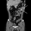
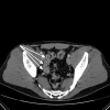
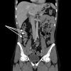
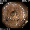
Idiopathic colonic varices represent a rare source of gastrointestinal haemorrhage with a presumed incidence around 0.0007%. Herein, we present a case of idiopathic colonic and small-intestine varices. According to our knowledge, this case report is the first description of both pan-colonic and small-intestine idiopathic varices of this extent. A young male patient without any previous notable medical history was admitted to the hospital because of massive enterorrhagia with haemodynamic instability. Colonoscopy revealed massive pan-colonic varices. After stabilization, numerous diagnostic procedures were performed in order to investigate the aetiology of pan-colonic varices without any explanation of the patient's condition. In addition, capsule endoscopy revealed varices through the whole length of the small intestine. The final diagnosis was idiopathic varices of the colon and small intestine. Because of the rapid clinical stabilization, the single incident of haemorrhage and the extension of the disease, a conservative approach was chosen (venotonics and β-blockers). During the 12-month follow-up period, the patient reported no gastrointestinal haemorrhage.
Keywords: Colon; Haemorrhage; Idiopathic colonic varices; Ileum; Varices.
Figures


References
-
- Šimeková K, Szilágyiová M, Antolová D, Laca Ľ, Polaček H, Nováková E, et al. Contribution to the Diagnosis and Treatment of Life-Threatening Parasitosis Caused by the Parasite Echinococcus multilocularis. Vector Borne Zoonotic Dis. 2017 Apr;17((4)):225–8. - PubMed
-
- Peixoto A, Silva M, Pereira P, Macedo G. Giant idiopathic pancolonic varices - a rare entity. Clin Res Hepatol Gastroenterol. 2016 Jun;40((3)):255–6. - PubMed
-
- Francois F, Tadros C, Diehl D. Pan-colonic varices and idiopathic portal hypertension. J Gastrointestin Liver Dis. 2007 Sep;16((3)):325–8. - PubMed
Publication types
LinkOut - more resources
Full Text Sources

