. 2019 Jan 9:11:492.
doi: 10.3389/fnmol.2018.00492. eCollection 2018.

Disruption of Otoferlin Alters the Mode of Exocytosis at the Mouse Inner Hair Cell Ribbon Synapse

Affiliations

Disruption of Otoferlin Alters the Mode of Exocytosis at the Mouse Inner Hair Cell Ribbon Synapse

Hideki Takago et al. Front Mol Neurosci. .

Abstract

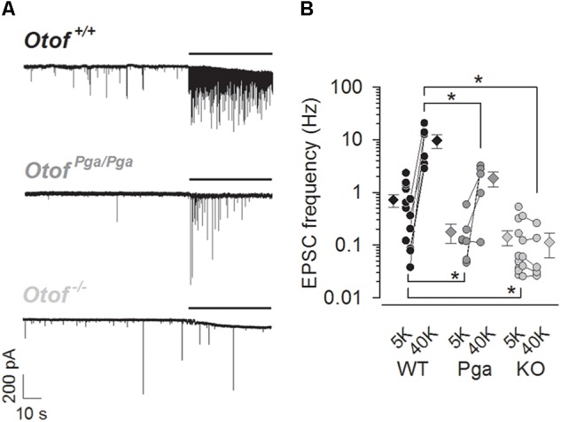
Sound encoding relies on Ca2+-mediated exocytosis at the ribbon synapse between cochlear inner hair cells (IHCs) and type I spiral ganglion neurons (SGNs). Otoferlin, a multi-C2 domain protein, is proposed to regulate Ca2+-triggered exocytosis at this synapse, but the precise mechanisms of otoferlin function remain to be elucidated. Here, performing whole-cell voltage-clamp recordings of excitatory postsynaptic currents (EPSCs) from SGNs in otoferlin mutant mice, we investigated the impact of Otof disruption at individual synapses with single release event resolution. Otof deletion decreased the spontaneous release rate and abolished the stimulus-secretion coupling. This was evident from failure of potassium-induced IHC depolarization to stimulate release and supports the proposed role of otoferlin in Ca2+ sensing for fusion. A missense mutation in the Otof gene (pachanga), in which otoferlin level at the IHC plasma membrane was lowered without changing its Ca2+ binding, also reduced the spontaneous release rate but spared the stimulus-secretion coupling. The slowed stimulated release rate supports the hypothesis that a sufficient abundance of otoferlin at the plasma membrane is crucial for the vesicle supply. Large-sized monophasic EPSCs remained present upon Otof deletion despite the drastic reduction of the rate of exocytosis. However, EPSC amplitude, on average, was modestly decreased. Moreover, a reduced contribution of multiphasic EPSC was observed in both Otof mutants. We argue that the presence of large monophasic EPSCs despite the exocytic defect upon Otof deletion supports the uniquantal hypothesis of transmitter release at the IHC ribbon synapse. Based upon the reduced contribution of multiphasic EPSC, we propose a role of otoferlin in regulating the mode of exocytosis in IHCs.

Keywords: EPSC; auditory; calcium; cochlea; hair cell; otoferlin; ribbon synapse; spiral ganglion neuron.


Figures

FIGURE 1
FIGURE 1
Otoferlin regulates spontaneous and high K+-stimulated release at the mouse IHC ribbon synapse. (A) Sample traces of EPSCs recorded from exemplar boutons of type I SGNs from a P9 Otof +/+ (top), a P8 OtofPga/Pga (middle) and a P9 Otof -/- (bottom) mouse. Each recording started in the extracellular solution containing 5.8 mM KCl, and 40 mM KCl was bath-applied during the time indicated by horizontal bars to depolarize presynaptic IHCs. (B) Summary of EPSC frequency in 5.8 mM (5K, spontaneous rate) and 40 mM (40 K, stimulated rate) [K+]e. Circles and diamonds indicate individual and mean ± SEM values for Otof +/+ (WT, black), OtofPga/Pga (Pga, dark gray) and Otof -/- (KO, light gray) SGNs. Asterisks at the bottom (spontaneous rate) and top (stimulated rate) show significant differences (p < 0.05). Note that the vertical axes for this and the next panels are shown on logarithmic scale. In some recordings, where 40 mM KCl was not applied, data points for 5.8 mM [K+]e alone were plotted.
FIGURE 2
FIGURE 2
Deletion of otoferlin does not abolish large EPSCs. Sample traces of monophasic (top row) and multiphasic (bottom rows) EPSCs recorded from Otof +/+ (left), Otof Pga/Pga (middle) and Otof -/- (right) SGNs.
FIGURE 3
FIGURE 3
Otoferlin deletion decreases EPSC amplitude and charge. (A–C) EPSC size for Otof +/+ (black), Otof Pga/Pga (dark gray) and Otof -/- (light gray) SGNs. Significant differences between Otof +/+ and Otof-/- SGNs (p < 0.05). Cumulative histograms of monophasic EPSC amplitude (A1), multiphasic EPSC amplitude (B1) and mono- and multiphasic (mixed) EPSC charge (C1) as well as individual (circles) and mean ± SEM (bars) values of monophasic EPSC amplitude (A2), multiphasic EPSC amplitude (B2) and mono- and multiphasic EPSC charge (C2) are shown. Note that a break line is inserted in the horizontal axis of panel C2 to clearly demonstrate the differences among groups.
FIGURE 4
FIGURE 4
Otoferlin disruption does not change EPSC kinetics (A,B) EPSC kinetics for Otof +/+ (black), Otof Pga/Pga (dark gray) and Otof -/- (light gray) SGNs. Individual (circles) and mean ± SEM (bars) values of 10–90 % rise time (A1) and decay time constant (A2) of monophasic EPSCs as well as time to peak (B1) and half-width (B2) of multiphasic EPSCs. No significant differences.
FIGURE 5
FIGURE 5
Otoferlin disruption decreases the fraction of multiphasic EPSCs (A,B) Exocytic mode of IHC exocytosis for Otof +/+ (black), Otof Pga/Pga (dark gray) and Otof -/- (light gray) SGNs. (A) Scatter plot of amplitude versus charge of monophasic (filled circles) and multiphasic (open circles) EPSCs derived from exemplar Otof +/+, OtofPga/Pga and Otof -/- SGNs. Note that the OtofPga/Pga and Otof -/- EPSCs cluster predominantly around the regression lines for monophasic EPSCs, which is indicative of less multiphasic EPSCs in Otof mutants than in Otof +/+. (B) Summary of the EPSC mode in 5.8 mM (5K, spontaneous rate) and 40 mM (40K, stimulated rate) [K+]e. Circles and diamonds indicate individual and mean ± SEM values of the percentage of multiphasic EPSCs for Otof +/+ (WT, black), OtofPga/Pga (Pga, dark gray) and Otof -/- (KO, light gray) SGNs. Asterisks at the bottom (spontaneous rate) and top (stimulated rate) show significant differences (∗∗p < 0.01 and ∗∗∗p < 0.001).

Similar articles

Cited by

References

    1. Beutner D., Moser T. (2001). The presynaptic function of mouse cochlear inner hair cells during development of hearing. J. Neurosci. 21 4593–4599. 10.1523/JNEUROSCI.21-13-04593.2001 - DOI - PMC - PubMed
    1. Chapochnikov N. M., Takago H., Huang C. H., Pangršič T., Khimich D., Neef J., et al. (2014). Uniquantal release through a dynamic fusion pore is a candidate mechanism of hair cell exocytosis. Neuron 83 1389–1403. 10.1016/j.neuron.2014.08.003 - DOI - PubMed
    1. Duncker S. V., Franz C., Kuhn S., Schulte U., Campanelli D., Brandt N., et al. (2013). Otoferlin couples to clathrin-mediated endocytosis in mature cochlear inner hair cells. J. Neurosci. 33 9508–9519. 10.1523/JNEUROSCI.5689-12.2013 - DOI - PMC - PubMed
    1. Glowatzki E., Fuchs P. A. (2002). Transmitter release at the hair cell ribbon synapse. Nat. Neurosci. 5 147–154. 10.1038/nn796 - DOI - PubMed
    1. Goodman M. B., Lockery S. R. (2000). Pressure polishing: a method for reshaping patch pipettes during fire polishing. J. Neurosci. Methods 100 13–15. 10.1016/S0165-0270(00)00224-7 - DOI - PubMed

LinkOut - more resources