Review
. 2019 Jan 11:11:493.
doi: 10.3389/fnmol.2018.00493. eCollection 2018.

Neuropeptides Exert Neuroprotective Effects in Alzheimer's Disease

Affiliations
Review

Neuropeptides Exert Neuroprotective Effects in Alzheimer's Disease

Xin-Yi Chen et al. Front Mol Neurosci. .

Abstract

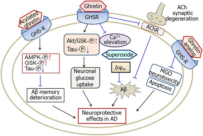
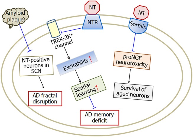
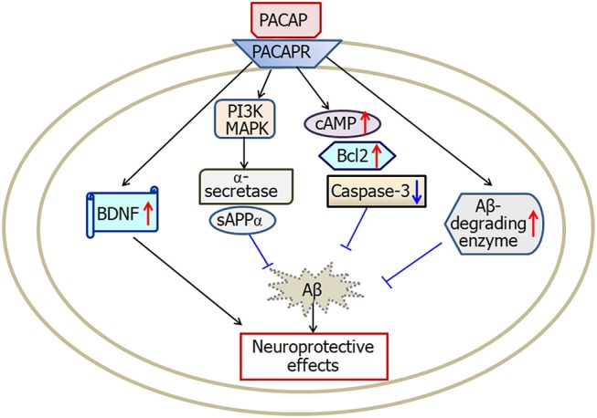
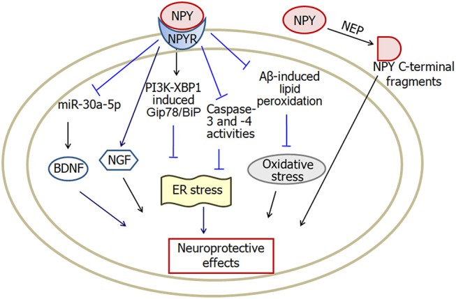
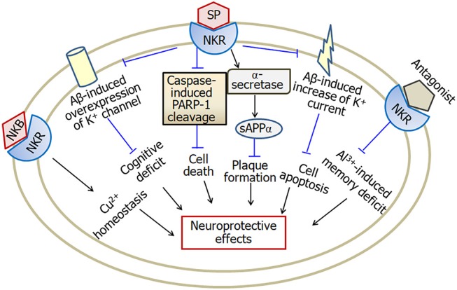
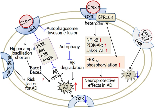
Alzheimer's disease (AD) is an age-related neurodegenerative disorder characterized by cognitive deficits and neuronal loss. Deposition of beta-amyloid peptide (Aβ) causes neurotoxicity through the formation of plaques in brains of Alzheimer's disease. Numerous studies have indicated that the neuropeptides including ghrelin, neurotensin, pituitary adenylate cyclase-activating polypeptide (PACAP), neuropeptide Y, substance P and orexin are closely related to the pathophysiology of Alzheimer's disease. The levels of neuropeptides and their receptors change in Alzheimer's disease. These neuropeptides exert neuroprotective roles mainly through preventing Aβ accumulation, increasing neuronal glucose transport, increasing the production of neurotrophins, inhibiting endoplasmic reticulum stress and autophagy, modulating potassium channel activity and hippocampal long-term potentiation. Therefore, the neuropeptides may function as potential drug targets in the prevention and cure of Alzheimer's disease.

Keywords: Alzheimer's disease; ghrelin; neuropeptide; neuropeptide Y; neurotensin; orexin; pituitary adenylate cyclase-activating polypeptide; substance P.


Figures

Figure 1
Figure 1
A schematic diagram describing the possible pathways of ghrelin-induced neuroprotective effects in Alzheimer's disease. Ghrelin protects against Aβ-induced neurotoxicity through prevention of calcium elevation, superoxide production and mitochondrial membrane depolarization. Ghrelin also increases neuronal glucose uptake by activating Akt/GSK phosphorylation and improving tau hyperphosphrylation. Moreover, ghrelin prevents cholinergic synaptic degeneration and therefore protects against Aβ-induced memory deficits. Acylated ghrelin improves Aβ-induced deterioration of memory through increase of AMPK and GSK phosphorylation and decrease of tau phosphorylation. Analog of ghrelin protects against MGO-induced neurotoxicity and apoptosis in cellular models of Alzheimer's disease. GHSR, growth hormone secretagogue receptors, also known as ghrelin receptors; Δψm, mitochondrial membrane potential; MGO, methylglyoxal; ACh, acetylcholine; AMPK, adenosine 5′-monophosphate (AMP)-activated protein kinase; GSK, glycogen synthase kinase; AChR, cholinergic receptors; -℗, phosphrylation. The internal and external circles represent the inner and outer leaflets of the cellular membrane. The dotted line in ACh synapse represents the degenerated synapse.
Figure 2
Figure 2
A model illustrating the neuroprotective effects of neurotensin in Alzheimer's disease. Neurotensin increases the excitability of neurons by inhibiting TREK-2K+ channel, which therefore improves memory status in Alzheimer's disease mice. Being a sortilin ligand, neurotensin rescues the survival of aged neurons through blocking sortilin-induced proNGF neurotoxicity. In addition, amyloid plaque density in the occipital cortex is negatively associated with neurotensin-positive neurons in the suprachiasmatic nucleus suggesting the involvement of neurotensin in fractal activity disruption in Alzheimer's disease. NT, neurotensin; NTR, neurotensin receptors; SCN, suprachiasmatic nucleus. The internal and external circles represent the inner and outer leaflets of the cellular membrane.
Figure 3
Figure 3
A scheme describing the possible mechanisms of PACAP-induced neuroprotective effects in Alzheimer's disease. PACAP protects against Aβ-induced neurotoxicity by activation of cAMP, BDNF, Bcl-2, Aβ-degrading enzyme and deactivation of caspase-3. Furthermore, PACAP increases α-secretase activation and then enhances secretion of sAPPα through both the MAPK and PI3K pathways. PACAP, pituitary adenylate cyclase-activating polypeptide; PACAPR, pituitary adenylate cyclase-activating polypeptide receptors; BDNF, brain-derived neurotrophic factor. The internal and external circles represent the inner and outer leaflets of the cellular membrane.
Figure 4
Figure 4
A model showing the possible pathways of neuropeptide Y-induced neuroprotective effects in Alzheimer's disease. Neuropeptide Y inhibits Aβ-induced lipid peroxidation and prevents intracellular oxidative stress. Activation of PI3K-XBP1 pathway may also be involved in neuropeptide Y-induced neuroprotection against endoplasmic reticulum stress. Moreover, both NGF and BDNF are involved in neuropeptide Y-induced neuroprotective effects. In addition, NEP cleaves neuropeptide Y into C-terminal fragments, which protect against the neurodegenerative pathology in Alzheimer's disease. NPY, neuropeptide Y; NPYR, neuropeptide Y receptors; NEP, neutral endopeptidase; ER, endoplasmic reticulum; BDNF, brain-derived neurotrophic factor; NGF, nerve growth factor. The internal and external circles represent the inner and outer leaflets of the cellular membrane.
Figure 5
Figure 5
A model describing the multiple effects of substance P in Alzheimer's disease. Substance P inhibits Aβ-induced overexpression of K+ channel and Aβ-induced increase of K+ current, and therefore attenuates cognitive deficit and apoptosis in Alzheimer's disease. Furthermore, substance P exerts neuroprotective effects through inhibition of caspase-3-induced PARP-1 cleavage and enhancement of α-secretase activity. Neurokinin B plays a role in copper homeostasis. However, substance P receptor antagonist attenuates aluminum-induced spatial memory deficit probably through blockade of substance P-mediated neuroinflammation. SP, substance P; NKR, neurokinin receptors; PARP-1, poly ADP-ribose polymerase-1; sAPPα, soluble amyloid precursor protein α; NKB, neurokinin B; Cu, copper; Al, aluminum. The internal and external circles represent the inner and outer leaflets of the cellular membrane.
Figure 6
Figure 6
A scheme describing the complicated effects of orexin in Alzheimer's disease. Firstly, in microglial cells, orexin suppresses autophagosome-lysosome fusion process, leading to impaired Aβ degradation. Furthermore, orexin suppresses Aβ uptake through downregulating phagocytosis regulating molecules, such as PI3K, Akt, and p38-MAPK. Aβ-plaque formation and tau hyper-phosphorylation decrease the expression of orexin receptors in Alzheimer's disease. Secondly, orexin receptors and GPR103 form functional heterodimers. Orexin augments NF-κB, PI3K-Akt, Jak-STAT signaling and induces ERK1/2 phosphorylation, and therefore is involved in neuroprotective functions. Finally, one of the orexin receptor 2 gene is likely a risk factor for Alzheimer's disease. In APP/PS1 Alzheimer's disease mice, orexin gene knock out decreases the amount of Aβ. Orexin modulates the hippocampal oscillation and the expression of clock-controlled-genes, Bace1 and Bace2, which are associated with the production of Aβ. OXR, orexin receptors; AD, Alzheimer's disease. The internal and external circles represent the inner and outer leaflets of the cellular membrane.

Similar articles

Cited by

References

    1. Ahmed M. M., Hoshino H., Chikuma T., Yamada M., Kato T. (2004). Effect of memantine on the levels of glial cells, neuropeptides, and peptide-degrading enzymes in rat brain regions of ibotenic acid-treated Alzheimer's disease model. Neuroscience 126, 639–649. 10.1016/j.neuroscience.2004.04.024 - DOI - PubMed
    1. Akbari E., Motamedi F., Naghdi N., Noorbakhshnia M. (2008). The effect of antagonization of orexin 1 receptors in CA1 and dentate gyrus regions on memory processing in passive avoidance task. Behav. Brain Res. 187, 172–177. 10.1016/j.bbr.2007.09.019 - DOI - PubMed
    1. Akbari E., Naghdi N., Motamedi F. (2006). Functional inactivation of orexin 1 receptors in CA1 region impairs acquisition, consolidation and retrieval in Morris water maze task. Behav. Brain Res. 173, 47–52. 10.1016/j.bbr.2006.05.028 - DOI - PubMed
    1. Al-Shawi R., Hafner A., Chun S., Raza S., Crutcher K., Thrasivoulou C., et al. . (2007). ProNGF, sortilin, and age-related neurodegeneration. Ann. N. Y. Acad. Sci. 1119, 208–215. 10.1196/annals.1404.024 - DOI - PubMed
    1. Al-Shawi R., Hafner A., Olsen J., Chun S., Raza S., Thrasivoulou C., et al. . (2008). Neurotoxic and neurotrophic roles of proNGF and the receptor sortilin in the adult and ageing nervous system. Eur. J. Neurosci. 27, 2103–2114. 10.1111/j.1460-9568.2008.06152.x - DOI - PubMed

LinkOut - more resources