

A 5-Year-Old Case of Choroidal Neovascularization in Enhanced S-Cone Syndrome Treated with Ranibizumab
- PMID: 30687072
- PMCID: PMC6341371
- DOI: 10.1159/000495743
A 5-Year-Old Case of Choroidal Neovascularization in Enhanced S-Cone Syndrome Treated with Ranibizumab
Abstract
Introduction: We describe the youngest case of enhanced S-cone syndrome (ESCS) associated with choroidal neovascularization (CNV) successfully treated with intravitreal ranibizumab injections.
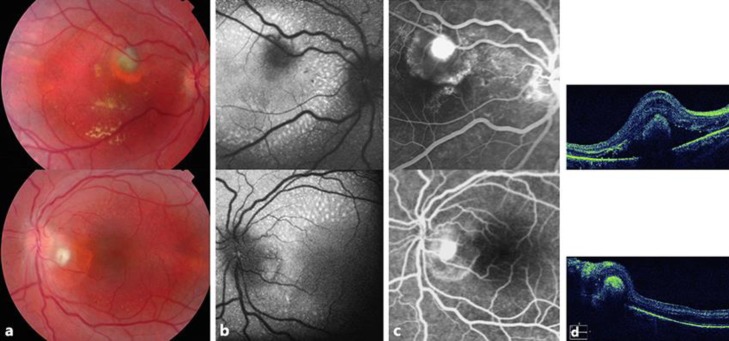
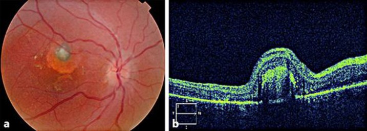
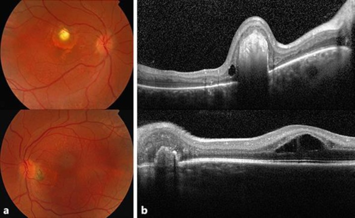
Case report: A 5-year-old boy presented with round-shaped fibrotic subretinal lesions in both eyes with surrounding subretinal fluid and progressive visual deterioration in the right eye. Fine foci of increased autofluorescence were observed along the arcades in both eyes. Fluorescein angiography revealed the presence of CNV in his right eye, and treatment with ranibizumab was initiated, with significant improvement in vision. Subsequent electroretinogram examination and genetic studies of the patient and his two younger siblings confirmed the diagnosis of ESCS.
Conclusion: CNV has been reported to occur in different inherited retinal degenerations, including ESCS. Our experience confirms that treatment with ranibizumab in patients with CNV-complicated ESCS can be potentially vision-saving.
Keywords: Anti-VEGF; Choroidal neovascularization; Electroretinogram; Enhanced S-cone syndrome; NR2E3; Ranibizumab.
Figures
References
-
- Hull S, Arno G, Sergouniotis PI, Tiffin P, Borman AD, Chandra A, et al. Clinical and molecular characterization of enhanced S-cone syndrome in children. JAMA Ophthalmol. 2014 Nov;132((11)):1341–9. - PubMed
-
- Schorderet DF, Escher P. NR2E3 mutations in enhanced S-cone sensitivity syndrome (ESCS), Goldmann-Favre syndrome (GFS), clumped pigmentary retinal degeneration (CPRD), and retinitis pigmentosa (RP) Hum Mutat. 2009 Nov;30((11)):1475–85. - PubMed
-
- Sustar M, Perovšek D, Cima I, Stirn-Kranjc B, Hawlina M, Brecelj J. Electroretinography and optical coherence tomography reveal abnormal post-photoreceptoral activity and altered retinal lamination in patients with enhanced S-cone syndrome. Doc Ophthalmol. 2015 Jun;130((3)):165–77. - PubMed
-
- Broadhead GK, Grigg JR, McCluskey P, Korsakova M, Chang AA. Bevacizumab for choroidal neovascularisation in enhanced S-cone syndrome. Doc Ophthalmol. 2016 Oct;133((2)):139–43. - PubMed
Publication types
LinkOut - more resources
Full Text Sources
