. 2019 Jan 9:9:1907.
doi: 10.3389/fphys.2018.01907. eCollection 2018.

Selenium-Containing Protein From Selenium-Enriched Spirulina platensis Attenuates Cisplatin-Induced Apoptosis in MC3T3-E1 Mouse Preosteoblast by Inhibiting Mitochondrial Dysfunction and ROS-Mediated Oxidative Damage

Affiliations

Selenium-Containing Protein From Selenium-Enriched Spirulina platensis Attenuates Cisplatin-Induced Apoptosis in MC3T3-E1 Mouse Preosteoblast by Inhibiting Mitochondrial Dysfunction and ROS-Mediated Oxidative Damage

Jing-Yi Sun et al. Front Physiol. .

Abstract

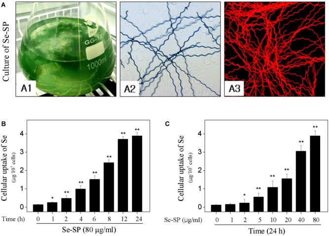
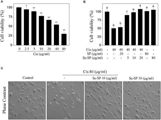
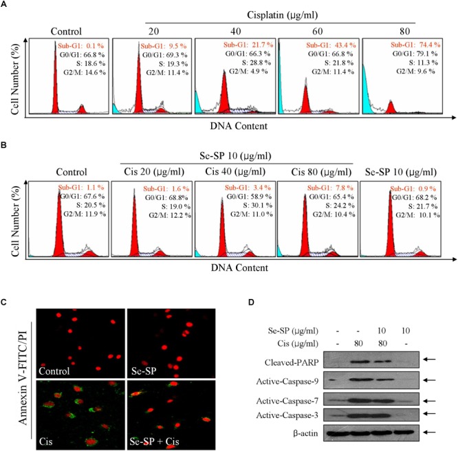
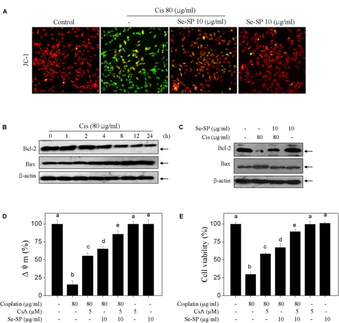
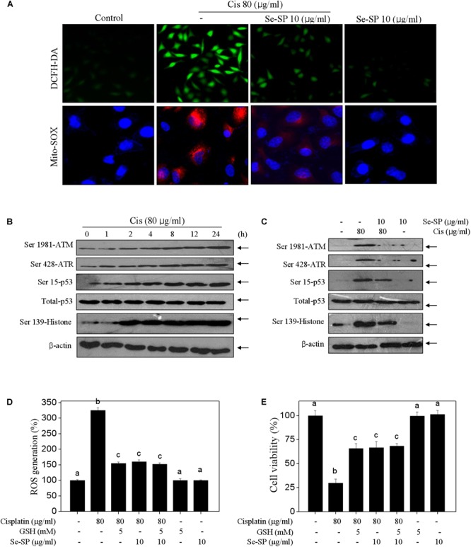
Accumulated evidences have verified that cancer chemotherapy may increase the risk of osteoporosis and severely affected the life quality. Osteoclasts hyperactivation was commonly accepted as the major pathogenesis of osteoporosis. However, the role of osteoblasts dysfunction in osteoporosis was little investigated. Our previous study has confirmed that selenium-containing protein from selenium-enriched Spirulina platensis (Se-SP) exhibited enhanced hepatoprotective potential through inhibiting oxidative damage. Herein, the protective effect of Se-SP against cisplatin-induced osteoblasts dysfunction in MC3T3-E1 mouse preosteoblast was investigated, and the underlying mechanism was evaluated. The results indicated that cisplatin dramatically decreased cell viability of preosteoblast by triggering mitochondria-mediated apoptosis pathway. Cisplatin treatment also caused mitochondrial dysfunction and reactive oxide species (ROS)-mediated oxidative damage. However, Se-SP pre-treatment effectively prevented MC3T3-E1 cells from cisplatin-induced mitochondrial dysfunction by balancing Bcl-2 family expression and regulating the opening of mitochondrial permeability transition pore (MPTP), attenuated cisplatin-induced oxidative damage through inhibiting the overproduction of ROS and superoxide anion, and eventually reversed cisplating-induced early and late apoptosis by inhibiting PARP cleavage and caspases activation. Our findings validated that Se-SP as a promising Se species could be a highly effective way in the chemoprevention and chemotherapy of oxidative damage-mediated bone diseases.

Keywords: apoptosis; cancer chemotherapy; mitochondrial dysfunction; osteoblasts dysfunction; osteoporosis; oxidative damage; selenium-containing protein.


Figures

FIGURE 1
FIGURE 1
Culture of Se-enriched Spirulina platensis and cellular uptake of Se. (A) Culture of Se-enriched S. platensis. (A1): Se-enriched S. platensis was cultured in 1000 ml Erlenmeyer flasks with Zarrouk medium. (A2): Phase contrast of Se-enriched S. platensis. (A3): Fluorescent morphology of Se-enriched S. platensis. Time-dependent (B) and dose-dependent (C) cellular uptake of Se. MC3T3-E1 cells were treated with 80 μg/ml Se-SP for 24 h, or cells were treated with 0–80 μg/ml Se-SP for 24 h. Cellular Se concentration was determined by ICP-AES method. All data and images were obtained from three independent experiments. Bars with “” or “∗∗” indicates P < 0.05 or P < 0.01, respectively. when compared with control group.
FIGURE 2
FIGURE 2
Selenium-SP inhibits cisplatin-induced cytotoxicity in MC3T3-E1 cells. (A) Dose-dependent cytotoxicity of cisplatin on MC3T3-E1 cells. Cells seeded in 96-well plate were treated with 0–80 μg/ml cisplatin for 24 h. (B) Se-SP inhibited cisplatin-induced cytotoxicity in MC3T3-E1 cells. Cells seeded in 96-well plate were pre-treated with 5–20 μg/ml SP or Se-SP for 24 h, and co-treated with 40 μg/ml cisplatin for another 24 h. Cell viability was detected by MTT assay. (C) Phase contrast of MC3T3-E1 cells morphology. Cells morphology was examined by phase-contrast microscopy. All data and images were obtained from three independent experiments. Bars with “” or “∗∗” indicates P < 0.05 or P < 0.01, respectively, when compared with control group. Bars with different characters are statistically different at P < 0.05 level, which achieved the multiple comparisons.
FIGURE 3
FIGURE 3
Selenium-SP reverses cisplatin-induced apoptosis in MC3T3-E1 cells. (A) Cisplatin-induced late apoptosis. Cells were treated with 20–80 μg/ml cisplatin for 24 h, and cells late apoptosis was examined by flow cytometric analysis. (B) Se-SP reversed cisplatin-induced late apoptosis in MC3T3-E1 cells. Cells were pre-treated Se-SP and co-treated with or without cisplatin. Then, Cells late apoptosis were analyzed by flow cytometric analysis. (C) Cells early apoptosis by annexin V-FITC/PI. Cells were pre-treated with 10 μg/ml Se-SP for 6 h, and/or co-treated with 80 μg/ml cisplatin for 6 h. Then, cells early apoptosis in living cells was imaged by annexin V-FITC/PI kit according to the manufacturer’s instructions. (D) Se-SP attenuated cisplatin-induced PARP cleavage and caspases activation. Cells seeded in 9-cm disk were pre-treated with 10 μg/ml Se-SP for 24 and/or co-treated with 80 μg/ml cisplatin for 24 h. Protein expression was assayed by western blotting method. All data and images were obtained from three independent experiments. Bars with different characters are statistically different at P < 0.05 level.
FIGURE 4
FIGURE 4
Selenium-SP blocks cisplatin-induced mitochondrial dysfunction. (A) Se-SP blocked cisplatin-induced loss of mitochondrial membrane potential (Δψm). MC3T3-E1 cells seeded in 6-well plate were pre-treated with 10 μg/ml Se-SP for 24 and/or co-treated with 80 μg/ml cisplatin for 24 h. Cells after treatment were labeled by JC-1 probe. The fluorescent shift from red to green was employed to indicate the loss of Δψm. (B) Time-dependent effect of cisplatin on Bcl-2 family. Cells were treated with 80 μg/ml cisplatin for 0–24 h. (C) Se-SP rescued cisplatin-induced Bcl-2 family imbalance. Bcl-2 and Bax were examined by western blotting. Effect of CsA on Δψm (D) and cell viability (E). Cells were pre-treated with 5 μM CsA for 2 h before combined treatment. Cell viability and Δψm were detected by MTT assay and JC-1 staining, respectively. All data and images were obtained from three independent experiments. Bars with different characters are statistically different at P < 0.05 level.
FIGURE 5
FIGURE 5
Selenium-SP attenuates ROS-mediated oxidative damage in cisplatin-treated MC3T3-E1 cells. (A) Se-SP prevented cisplatin-induced accumulation of ROS and superoxide production. MC3T3-E1 cells seeded in 6-well plate were pre-treated with 10 μg/ml Se-SP for 24 h and/or co-treated with 80 μg/ml cisplatin for 24 h. Cells after treatment were labeled by DCFH-DA (green) or Mito-SOX (red) for detection of ROS and superoxide production, respectively. (B) Time-dependent effect of cisplatin on DNA damage signal axis. Cells were treated with 80 μg/ml cisplatin for 0–24 h. (C) Se-SP attenuated cisplatin-induced DNA damage. Protein expression was examined by western blotting. Effect of GSH on ROS generation (D) and cell viability (E). Cells were pre-treated with 5 mM GSH for 2 h before combined treatment. Cell viability and ROS generation were detected by MTT assay and DCFH-DA staining, respectively. All data and images were obtained from three independent experiments. Bars with different characters are statistically different at P < 0.05 level.

References

    1. Bashandy S. A., El Awdan A. S., Ebaid H., Alhazza I. M. (2016). Antioxidant potential of Spirulina platensis mitigates oxidative stress and reprotoxicity induced by sodium arsenite in male rats. Oxid. Med. Cell. Longev. 2016:7174351. 10.1155/2016/7174351 - DOI - PMC - PubMed
    1. Brecht K., Simonen M., Heim J. (2005). Upregulation of alpha globin promotes apoptotic cell death in the hematopoietic cell line FL5.12. Apoptosis 10 1043–1062. 10.1007/s10495-005-0617-9 - DOI - PubMed
    1. Brown K. M., Arthur J. R. (2001). Selenium, selenoproteins and human health: a review. Public Health Nutr. 4 593–599. 10.1079/PHN2001143 - DOI - PubMed
    1. Bruijnincx P. C., Sadler P. J. (2008). New trends for metal complexes with anticancer activity. Curr. Opin. Chem. Biol. 12 197–206. 10.1016/j.cbpa.2007.11.013 - DOI - PMC - PubMed
    1. Cervellati C., Bonaccorsi G., Cremonini E., BergaMini C. M., Patella A., Castaldini C., et al. (2013). Bone mass density selectively correlates with serum markers of oxidative damage in post-menopausal women. Clin. Chem. Lab. Med. 51 333–338. 10.1515/cclm-2012-0095 - DOI - PubMed

LinkOut - more resources